. 2024 Nov 13;14(11):548.
doi: 10.3390/bios14110548.

Enhanced Detection of Vibrio harveyi Using a Dual-Composite DNAzyme-Based Biosensor

Affiliations

Enhanced Detection of Vibrio harveyi Using a Dual-Composite DNAzyme-Based Biosensor

Siying Li et al. Biosensors (Basel). .

Abstract

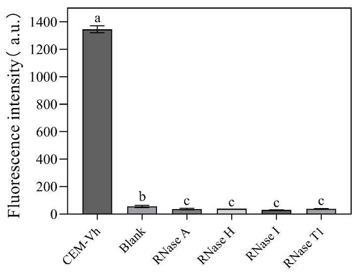
Vibrio harveyi is a serious bacterial pathogen which can infect a wide range of marine organisms, such as marine fish, invertebrates, and shrimp, in aquaculture, causing severe losses. In addition, V. harveyi can be transmitted through food and water, infecting humans and posing a serious threat to public safety. Therefore, rapid and accurate detection of this pathogen is key for the prevention and control of related diseases. In this study, nine rounds of in vitro screening were conducted with Systematic Evolution of Ligands by Exponential Enrichment (SELEX) technology using unmodified DNA libraries, targeting the crude extracellular matrix (CEM) of V. harveyi. Two DNAzymes, named DVh1 and DVh3, with high activity and specificity were obtained. Furthermore, a fluorescent biosensor with dual DNAzymes was constructed which exhibited improved detection efficiency. The sensor showed a good fluorescence response to multiple aquatic products (i.e., fish, shrimp, and shellfish) infected with V. harveyi, with a detection limit below 11 CFU/mL. The fluorescence signal was observed within 30 min of reaction after target addition. This simple, inexpensive, highly effective, and easy to operate DNAzymes biosensor can be used for field detection of V. harveyi.

Keywords: SELEX; Vibrio harveyi; dual DNAzyme; field detection.


Conflict of interest statement

The authors confirm that there is no conflict of interests regarding this paper.

Figures

Figure 1
Figure 1
Flowchart of DNAzyme screening. The library contained 40 random nucleotides and was screened for nine rounds. Negative selection was carried out during the 5th and 7th rounds of screening, while positive selections were conducted in the other rounds. Biotin was labeled at the 5′ end. The target molecule was the CEM of bacteria.
Figure 2
Figure 2
Activity of candidate DNAzymes: (a) fluorescence intensity (DVh1–6 reacted with CEM-Vh); and (b) results of candidate 15% dPAGE (DVh1–6 reacted with CEM-Vh, Blank1–6 mean DVh1–6 reacted without CEM-Vh).
Figure 3
Figure 3
Differences between the fluorescence intensity signals of single and dual DNAzymes. (Blank means without adding CEM-Vh, CEM-Vh means adding CEM-Vh. Different letters indicate statistically significant differences (p < 0.05), while identical letters indicate insignificant differences (p > 0.05)).
Figure 4
Figure 4
Optimization of reaction conditions: (a) pH optimization; (b) influence of various divalent metal ions on the cleavage activity of DVh3+1 (different letters indicate statistically significant differences (p < 0.05), while identical letters indicate insignificant differences (p > 0.05)); and (c) optimization of the concentrations of Na+ and Mg2+. Buffer/EDTA reaction contained 300 mM EDTA in 2× selection buffer. The bars and the dots represent mean ± SD.
Figure 5
Figure 5
Specificity of DVh3+1: (a) fluorescence intensity of DVh3+1 in presence of CEM of various bacteria; (b) specificity of DVh3+1 analyzed by 15% dPAGE (Blank: reaction system without CEM-Vh).
Figure 6
Figure 6
Sensitivity of DVh3+1: (a) fluorescence intensity signals of different concentrations of V. harveyi (Blank: normal saline) and the calibration curves constructed using the fluorescence signals corresponding to 4.7 × 101, 4.7 × 10², and 4.7 × 10³ CFU/mL of V. harveyi; and (b) gel cleavage assay at different concentrations of V. harveyi (Blank: reaction system without CEM-Vh).
Figure 7
Figure 7
Identification of the target of DVh3+1: (a) fluorescence intensity of untreated and protease-treated CEM-Vh cleaved by DVh3+1; (b) 15% dPAGE analysis of the cleavage activity of DVh3+1 against CEM-Vh treated with various proteases; (c) fluorescence intensity of the reactions between DVh3+1 and CEM-Vh with different molecular weights; (d) 15% dPAGE analysis of the cleavage activity of DVh3+1 against CEM-Vh with different molecular weights. (Blank: reaction system without CEM-Vh).
Figure 8
Figure 8
Effect of four different RNases on the cleavage activity of DVh3+1 (Blank: reaction system without CEM-Vh; Different letters indicate statistically significant differences (p < 0.05), while identical letters indicate insignificant differences (p > 0.05)).
Figure 9
Figure 9
(a) Optimization of the concentration of DVh3+1-S in the dual DNAzyme sensor, with corresponding pictures of fluorescence signal shown at the top. (b) Analysis of significant differences between the fluorescence values of different concentrations of DVh3+1-S at different reaction times. Different letters indicate statistically significant differences (p < 0.05), while the same letters indicate insignificant differences (p > 0.05). The bar represents the mean ± SD.
Figure 10
Figure 10
Comparisons of the aquatic products infected by V. harveyi: (a) P. vannamei before infection, (b) P. vannamei after infection 24 h, (c) whelk before infection, (d) Whelk after infection 60 h, (e) C. formosana before infection, (f) C. formosana after infection 48 h, (g) Epinephelus before infection and (h) Epinephelus after infection 50 h.
Figure 11
Figure 11
V. harveyi detection in the actual samples by DVh3+1 sensor: (a) fluorescence intensities of blank and four actual samples (C. formosana, Epinephelus, P. vannamei, and whelk; Blank: uninfected animal samples); (b) fluorescence intensities of diluted whelk samples, with calibration curves plotted using the fluorescence values of V. harveyi at concentrations of 1.02 × 101, 1.02 × 10², and 1.02 × 10³ CFU/mL (Different letters indicate statistically significant differences (p < 0.05)).

Similar articles

References

    1. Nguyen H.T., Thu Nguyen T.T., Tsai M.A., Ya-Zhen E., Wang P.C., Chen S.C. A formalin-inactivated vaccine provides good protection against Vibrio harveyi infection in orange-spotted grouper (Epinephelus coioides) Fish Shellfish Immunol. 2017;65:118–126. doi: 10.1016/j.fsi.2017.04.008. - DOI - PubMed
    1. Montanchez I., Kaberdin V.R. Vibrio harveyi A brief survey of general characteristics and recent epidemiological traits associated with climate change. Mar. Environ. Res. 2020;154:104850. doi: 10.1016/j.marenvres.2019.104850. - DOI - PubMed
    1. Zhang X.-H., He X., Austin B. Vibrio harveyi: A serious pathogen of fish and invertebrates in mariculture. Mar. Life Sci. Technol. 2020;2:231–245. doi: 10.1007/s42995-020-00037-z. - DOI - PMC - PubMed
    1. Gan L., Zheng J., Xu W.-H., Lin J., Liu J., Zhang Y., Wu Z., Lv Z., Jia Y., Guo Q., et al. Deciphering the virulent Vibrio harveyi causing spoilage in muscle of aquatic crustacean Litopenaeus vannamei. Sci. Rep. 2022;12:16296. doi: 10.1038/s41598-022-20565-1. - DOI - PMC - PubMed
    1. Dubert J., Nelson D.R., Spinard E.J., Kessner L., Gomez-Chiarri M., Costa F.d., Prado S., Barja J.L. Following the infection process of vibriosis in Manila clam (Ruditapes philippinarum) larvae through GFP-tagged pathogenic Vibrio species. J. Invertebr. Pathol. 2016;133:27–33. doi: 10.1016/j.jip.2015.11.008. - DOI - PubMed

Supplementary concepts

LinkOut - more resources