. 2024 Oct 30;12(11):347.
doi: 10.3390/dj12110347.

Predictable Full Digital Workflow Using Stackable Surgical Templates for Complete Dental Arch Rehabilitation with Implant-Supported Fixed Restorations-Case Series and Proof of Concept

Affiliations

Predictable Full Digital Workflow Using Stackable Surgical Templates for Complete Dental Arch Rehabilitation with Implant-Supported Fixed Restorations-Case Series and Proof of Concept

Corina Marilena Cristache et al. Dent J (Basel). .

Abstract

Background: In recent years, advancements in digital dentistry have provided new opportunities for more predictable and efficient treatment options, particularly in patients with failing dentition. This study aimed to evaluate the effectiveness and accuracy of a fully digital workflow using stackable surgical templates for complete dental arch rehabilitation with implant-supported fixed restorations.

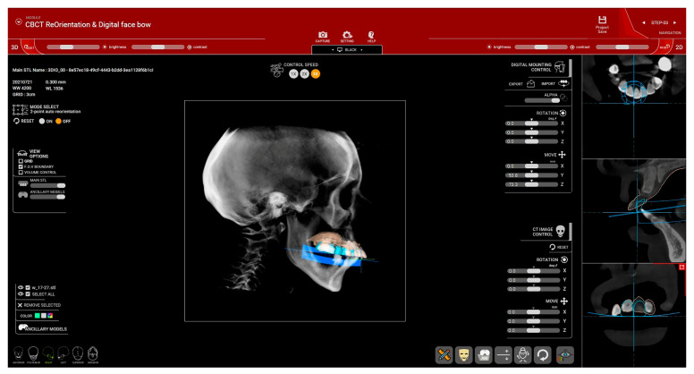
Methods: Four patients, comprising two males and two females with a mean age of 66 years, were included in this case series. Each patient underwent meticulous digital planning, including CBCT and intraoral scanning, to create a virtual patient for preoperative assessment and virtual treatment planning. The assessment of the trueness of implant positioning was conducted in Geomagic Control X software (version 2017.0.3) by referencing anatomical landmarks from both the preoperative and one-year postoperative CBCT scans.

Results: A total of 25 dental implants were placed in the maxilla, followed by the installation of long-term provisional restorations. The results showed minimal deviation between the planned and actual implant positions, with mean 3D coronal, apical, and angular discrepancies of 0.87 mm, 2.04 mm, and 2.67°, respectively. All implants achieved successful osseointegration, and no failures were recorded, resulting in a 100% survival rate at the one-year follow-up. Patients reported high satisfaction with both the esthetic and functional outcomes based on their subjective feedback.

Conclusions: The findings suggest that the use of a fully digital workflow with stackable surgical templates is a reliable and effective approach for immediate implant placement and prosthetic rehabilitation, enhancing treatment precision and patient comfort.

Keywords: edentulous maxilla; full digital workflow; immediate fixed prosthesis; stackable guides; stackable surgical template; virtual patient.


Conflict of interest statement

The authors declare no conflicts of interest.

Figures

Figure 1
Figure 1
Overview of the steps involved in using R2Gate™ software for virtual patient creation and treatment planning.
Figure 2
Figure 2
CBCT re-orientation (Patient #1): (a) frontal alignment of CBCT—centering and orienting with the orbital line parallel to the horizontal line; (b) lateral view with the Frankfort plane set as the horizontal plane.
Figure 3
Figure 3
(Patient#1): (a) DICOM files from the CBCT, STL files from the intraoral scan (edentulous maxilla with preexisting adapted denture), and OBJ files from facial scanning were imported into the software; (b) fine manual alignment of the DICOM and STL files.
Figure 4
Figure 4
(Patient #1): Cephalometric analysis of the anteroposterior (AP) position of the maxilla (Mx) and mandible (Mn) for determining the A-Nasion-B (ANB) angle. Cephalometriclandmarks are displayed in the image on the right. N = nasion, S = sella, Or = orbitale, A = subspinale, UIA = upper incisor root apex, UIT = upper incisor incisal edge, LIT = lower incisor incisal edge, LIA = lower incisor root apex, B = supramentale, Pog = pogonion.
Figure 5
Figure 5
(Patient #2): Kim’s cephalometric analysis for assessing the positioning of the maxilla in relation to the McNamara line, as well as evaluating the Y-angle and occlusal plane (OP) angle for precise treatment planning. N = Nasion, S = Sella, Or = Orbitale, Po = Porion, A = Point A, 6 = the root apex of the upper incisor, 7 = the incisal edge of the upper incisor, 8 = the posterior point of occlusion, Pog = Pogonion, Me = Menton, Go = Gonion.
Figure 6
Figure 6
(Patient #2): Digital Mounting in the Articulator: (a) Artex articulator with the Frankfort horizontal plane selected as the reference plane in R2Gate™ software; (b) mounting imported as an STL file into EXOCAD software for the design of the provisional restoration.
Figure 7
Figure 7
(Patient #2): Occlusal plane alignment using R2Gate™ software.
Figure 8
Figure 8
(Patient #2): Digital Smile Design in EXOCAD software for selecting anterior teeth, followed by designing the provisional restoration based on previously obtained information.
Figure 9
Figure 9
(Patient #1): Prosthetically driven implant planning. Six implants were planned for this patient, along with the design of four transverse pins for base guide stabilization.
Figure 10
Figure 10
(Patient #2): Detailed positioning of the implants and transversal pins generated by R2Gate™ software according to the surgical plan. Bone density and the recommended drilling sequence for each implant position are also displayed. The R2Gate™ software converts the CBCT grayscale into five basic colors corresponding to the 256 shades of gray (right images): black represents air, red indicates soft tissue, blue represents soft bone, yellow corresponds to dense bone, and green represents high-density structures (such as enamel, cortical bone, and metal structures).
Figure 11
Figure 11
(Patient #3): Design of stackable guides (from left to right): maxillary model before extractions, base guide, tooth-supported guide for the insertion of the first two implants; model with virtual extractions, base guide, implant-supported guide; model with virtual extractions, base guide, and prosthetic guide.
Figure 12
Figure 12
Matrix (displayed in blue)-patrix (displayed in beige) type of attachment between the base guide and subsequent surgical or prosthetic guides.
Figure 13
Figure 13
3D Printed Stackable Guides: (a) base guide on the 3D-printed model with implant digital analogs and OT Equator™ corresponding abutments in the planned positions; (b) mucosa-supported guide for dental implant insertion; (c) prosthetic guide with temporary fixed restoration; (d) temporary fixed restoration with temporary cylinders in place and Seeger™ conical Teflon rings (OT Equator™ abutments with temporary cylinders and Seeger™ conical Teflon rings are also displayed separately); (e) base guide and mucosa-supported guide with putty bite for base guide fixation using transverse pins.
Figure 14
Figure 14
Aligned preoperative CBCT with treatment plan (right) and postoperative CBCT (left) in R2Gate software.
Figure 15
Figure 15
Assessment of the 3D positions of the planned implants (shown in blue) versus the placed implants (shown in green). Tolerance limits were set to ±0.1 mm (indicated by green on the scale). The 3D deviation for each point was calculated as the square root of the sum of the squared deviations along the three axes (x, y, and z).

References

    1. Papaspyridakos P., Bedrossian A., De Souza A., Bokhary A., Gonzaga L., Chochlidakis K. Digital Workflow in Implant Treatment Planning for Terminal Dentition Patients. J. Prosthodont. 2022;31:543–548. doi: 10.1111/jopr.13510. - DOI - PubMed
    1. Coachman C., Calamita M.A., Coachman F.G., Coachman R.G., Sesma N. Facially generated and cephalometric guided 3D digital design for complete mouth implant rehabilitation: A clinical report. J. Prosthet. Dent. 2017;117:577–586. doi: 10.1016/j.prosdent.2016.09.005. - DOI - PubMed
    1. Ghionea I.G., Vatamanu O.E.B., Cristescu A.M., David M., Stancu I.C., Butnarasu C., Cristache C.M. A Finite Element Analysis of a Tooth-Supported 3D-Printed Surgical Guide without Metallic Sleeves for Dental Implant Insertion. Appl. Sci. 2023;13:9975. doi: 10.3390/app13179975. - DOI
    1. Chandran S., Sers L., Picciocchi G., Luongo F., Lerner H., Engelschalk M., Omar S. Guided implant surgery with R2Gate®: A multicenter retrospective clinical study with 1 year of follow-up. J. Dent. 2022;127:104349. doi: 10.1016/j.jdent.2022.104349. - DOI - PubMed
    1. Cristache C.M., Burlibasa M., Tudor I., Totu E.E., Di Francesco F., Moraru L. Accuracy, Labor-Time and Patient-Reported Outcomes with Partially versus Fully Digital Workflow for Flapless Guided Dental Implants Insertion—A Randomized Clinical Trial with One-Year Follow-Up. J. Clin. Med. 2021;10:1102. doi: 10.3390/jcm10051102. - DOI - PMC - PubMed

LinkOut - more resources