. 2024 Oct 24;14(11):1071.
doi: 10.3390/jpm14111071.

The Three-Dimensional Investigation of the Radiographic Boundary of Mandibular Full-Arch Distalization in Different Facial Patterns

Affiliations

The Three-Dimensional Investigation of the Radiographic Boundary of Mandibular Full-Arch Distalization in Different Facial Patterns

Yin-Yu Chou et al. J Pers Med. .

Abstract

Objective: Mandibular full-arch distalization (MFD) is a popular approach, particularly in non-extraction cases. However, we still cannot confirm whether facial patterns affect the amount of limits. This study aimed to determine the anatomical MFD limits in patients with different facial patterns.

Study design: Using computed tomography (CT), the shortest distances from the mandibular second molar to the inner cortex of the mandibular lingual surface and from the lower central incisor to the inner cortex of the lingual mandibular symphysis were measured in 60 samples (30 patients). The available distalization space in both regions was compared between groups with different facial patterns.

Results: The available space in symphysis was more critical than that in retromolar area: the shortest distances to the inner cortex of the lingual mandibular symphysis at root levels 8 mm apical to the cementoenamel junction of the incisor were 1.28, 1.60, and 3.48 mm in the high-, normal-, and low-angle groups, respectively.

Conclusions: Facial patterns affected the MFD capacity, and the thickness of the lingual mandibular symphysis was the most critical anatomic limit encountered. Practitioners should always pay attention to the possible impacts from facial patterns, especially in the treatment of high-angle cases.

Keywords: computed tomography; facial pattern; full-arch distalization; mandible.


Conflict of interest statement

The authors declare no conflicts of interest.

Figures

Figure 1
Figure 1
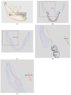
Reconstructed mandibular model with reference index. Blue dots: the three reference points constituting the mandibular occlusal plane. (a) Posterior lingual boundary: the shortest distance posterior to the mandibular second molar (dotted red line: the posterior occlusal line connecting the most occlusal point of the buccal grooves of the mandibular first and second molars). (b) Thickness of the lingual mandibular symphysis: the shortest distance lingual to the mandibular central incisor (dotted green line: the line perpendicular to the incisal edge from the occlusal view).
Figure 2
Figure 2
Posterior lingual boundary (in mm). (A). The axial slices at the depths of the crown-top level (solid gray line) and 2, 4, 6, and 8 mm apical to the CEJ (dotted gray lines) were made parallel to the designated mandibular occlusal plane. (B). The shortest distance from the mandibular second molar at the crown-top level to the outer cortex of the anterior surface of ramus (PCr-0 mm). (solid red line: the parallel index to the posterior occlusal line). (C). The shortest distance from the most lingual point of the distal root of the mandibular second molar to the inner cortex of the mandibular lingual surface at 2, 4, 6, and 8 mm apical to the CEJ (PRoin-2, -4, -6, and -8 mm, respectively). (solid red line: the parallel index to the posterior occlusal line) (D). Close-up view of the black box in B, showing the shortest distance to the outer cortex of the anterior surface of ramus (PCr-0 mm). (E). Close-up view of the black box in C, showing the shortest distance to the inner cortex of the mandibular lingual surface (PRoin-2, -4, -6, and -8 mm).
Figure 3
Figure 3
Thickness of the lingual mandibular symphysis (in mm). (A). The axial slices at depths of 0, 2, 4, 6, and 8 mm apical to the CEJ (dotted white lines) were made parallel to the designated mandibular occlusal plane. (B). Measurements of the shortest distance from the lingual point of the mandibular central incisor root to the inner cortex of the lingual mandibular symphysis at depths of 0, 2, 4, 6, and 8 mm apical to the CEJ on axial slices (ARoin-0, -2, -4, -6, and -8 mm, respectively). (solid green line: the parallel index perpendicular to the incisal edge of the mandibular central incisor). (C). Close-up view of the black box in B, showing the shortest distance to the inner cortex of the lingual mandibular symphysis (ARoin-0, -2, -4, -6, and -8 mm).
Figure 3
Figure 3
Thickness of the lingual mandibular symphysis (in mm). (A). The axial slices at depths of 0, 2, 4, 6, and 8 mm apical to the CEJ (dotted white lines) were made parallel to the designated mandibular occlusal plane. (B). Measurements of the shortest distance from the lingual point of the mandibular central incisor root to the inner cortex of the lingual mandibular symphysis at depths of 0, 2, 4, 6, and 8 mm apical to the CEJ on axial slices (ARoin-0, -2, -4, -6, and -8 mm, respectively). (solid green line: the parallel index perpendicular to the incisal edge of the mandibular central incisor). (C). Close-up view of the black box in B, showing the shortest distance to the inner cortex of the lingual mandibular symphysis (ARoin-0, -2, -4, -6, and -8 mm).
Figure 4
Figure 4
A simplified diagram summarizing the thinking process to evaluate the treatment strategies in patients of Class III malocclusion among different facial patterns.

References

    1. Ye J.-A., Tsai C.-Y., Lee Y.-H., Chang Y.-J., Lin S.-S., Lai J.-P., Wu T.-J. Could cephalometric landmarks serve as boundaries of maxillary molar distalization? A comparison between two-and three-dimensional assessments. Taiwan. J. Orthod. 2021;33:1. doi: 10.38209/2708-2636.1105. - DOI
    1. López D.F., Olmos D.A., Morales M.A. Anatomical factors of the maxillary tuberosity that influence molar distalization. Korean J. Orthod. 2024;54:239–246. doi: 10.4041/kjod24.017. - DOI - PMC - PubMed
    1. Park J.H., Kook Y.-A., Kim Y.J., Lee N.-K. Biomechanical considerations for total distalization of the maxillary dentition using TSADs. Semin. Orthod. 2020;26:139–147. doi: 10.1053/j.sodo.2020.06.011. - DOI
    1. Kim S.-H., Cha K.-S., Lee J.-W., Lee S.-M. Mandibular skeletal posterior anatomic limit for molar distalization in patients with Class III malocclusion with different vertical facial patterns. Korean J. Orthod. 2021;51:250. doi: 10.4041/kjod.2021.51.4.250. - DOI - PMC - PubMed
    1. Kim S.-J., Choi T.-H., Baik H.-S., Park Y.-C., Lee K.-J. Mandibular posterior anatomic limit for molar distalization. Am. J. Orthod. Dentofac. Orthop. 2014;146:190–197. doi: 10.1016/j.ajodo.2014.04.021. - DOI - PubMed

LinkOut - more resources