. 2024 Oct 26;10(11):741.
doi: 10.3390/jof10110741.

Label-Free Optical Transmission Tomography for Direct Mycological Examination and Monitoring of Intracellular Dynamics

Affiliations

Label-Free Optical Transmission Tomography for Direct Mycological Examination and Monitoring of Intracellular Dynamics

Eliott Teston et al. J Fungi (Basel). .

Abstract

Live-cell imaging generally requires pretreatment with fluorophores to either monitor cellular functions or the dynamics of intracellular processes and structures. We have recently introduced full-field optical coherence tomography for the label-free live-cell imaging of fungi with potential clinical applications for the diagnosis of invasive fungal mold infections. While both the spatial resolution and technical set up of this technology are more likely designed for the histopathological analysis of tissue biopsies, there is to our knowledge no previous work reporting the use of a light interference-based optical technique for direct mycological examination and monitoring of intracellular processes. We describe the first application of dynamic full-field optical transmission tomography (D-FF-OTT) to achieve both high-resolution and live-cell imaging of fungi. First, D-FF-OTT allowed for the precise examination and identification of several elementary structures within a selection of fungal species commonly known to be responsible for invasive fungal infections such as Candida albicans, Aspergillus fumigatus, or Rhizopus arrhizus. Furthermore, D-FF-OTT revealed the intracellular trafficking of organelles and vesicles related to metabolic processes of living fungi, thus opening new perspectives in fast fungal infection diagnostics.

Keywords: fungal metabolism; invasive fungal infections; live-cell imaging; medical mycology; optical tomography.


Conflict of interest statement

The authors declare no conflicts of interest.

Figures
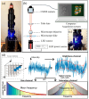
Figure 1
Figure 1
Illustration (a) and schematic representation of the prototype OTT system (b), focus on the objective area of the microscope (c), and a schematic explanation of signal treatment for dynamic imaging reconstruction (d) during Candida albicans imaging. The computer window in (b) shows both structural greyscale (left) and dynamic (right) modes. (Adapted from Ref [17]).
Figure 2
Figure 2
Bright-field and D-FF-OTT imaging of Candida albicans (ad) and Candida parapsilosis (eh). Bright-field imaging of Candida albicans (a) and Candida parapsilosis (e). FF-OTT (b,f) and D-FF-OTT (c,d,g,h) of Candida albicans (upper row) and parapsilosis (lower row). Dashed squares in Figure 2 (c,g) are shown with a higher magnification in (d,h). White arrows indicate cell membranes, empty triangles indicate nuclear membranes, and white-filled arrowheads point out organelles. Scale bars represent 10 μm.
Figure 3
Figure 3
Bright-field and D-FF-OTT imaging of Aspergillus fumigatus. Bright-field imaging of unstained (a,e) and lactophenol blue-stained (b,f) fungi. FF-OTT (c,g) and D-FF-OTT (d,h) of similar structures. White arrowheads point out septa. Scale bars represent 10 μm.
Figure 4
Figure 4
Bright-field and FF-OTT imaging of Aspergillus niger. Bright-field imaging of unstained (a,e) and lactophenol blue-stained (b,f) fungi. FF-OTT (c,g) and D-FF-OTT (d,h) of similar structures. White arrowheads point out septa. Scale bars represent 10 μm.
Figure 5
Figure 5
Optical and FF-OTT imaging of Rhizopus arrhizus. Optical imaging of unstained (a,e) and lactophenol blue-stained (b,f) fungi. FF-OTT (c,g) and D-FF-OTT (d,h) of similar structures. White arrowheads indicate sporangiospores. Scale bars represent 10 μm.
Figure 6
Figure 6
Quantitative spectral analysis of cellular structure of Candida albicans. D-FF-OTT registers three spectral parameters that are the central fluctuation frequency (a,b), frequency range (c,d), and amplitude of fluctuations (e,f). Images were analyzed and signals were quantified for each spectral characteristic for three distinct structures, namely the plasma membranes (blue circles), nuclear membranes (orange squares), and lipid droplets (purple triangles). Statistical analysis showed a significant difference (p < 0.0001) for any comparison except for the comparison between the amplitude of fluctuations of plasma and nuclear membranes, as indicated on corresponding graph (ns in (f)). White arrows indicate cell membranes, empty triangles indicate nuclear membranes, and white-filled arrowheads point out organelles. Scale bars represent 10 μm.

References

    1. Brown G.D., Denning D.W., Gow N.A., Levitz S.M., Netea M.G., White T.C. Hidden Killers: Human Fungal Infections. Sci. Transl. Med. 2012;4:165rv13. doi: 10.1126/scitranslmed.3004404. - DOI - PubMed
    1. Denning D.W. Global incidence and mortality of severe fungal disease. Lancet Infect. Dis. 2024;24:e428–e438. doi: 10.1016/S1473-3099(23)00692-8. - DOI - PubMed
    1. Danion F., Rouzaud C., Duréault A., Poirée S., Bougnoux M.E., Alanio A., Lanternier F., Lortholary O. Why are so many cases of invasive aspergillosis missed? Med. Mycol. 2019;57:S94–S103. doi: 10.1093/mmy/myy081. - DOI - PubMed
    1. Vallabhaneni S., Mody R.K., Walker T., Chiller T. The Global Burden of Fungal Diseases. Infect. Dis. Clin. N. Am. 2016;30:1–11. doi: 10.1016/j.idc.2015.10.004. - DOI - PubMed
    1. Tochigi N., Sadamoto S., Oura S., Kurose Y., Miyazaki Y., Shibuya K. Artificial Intelligence in the Diagnosis of Invasive Mold Infection: Development of an Automated Histologic Identification System to Distinguish Between Aspergillus and Mucorales. Med. Mycol. J. 2022;63:91–97. doi: 10.3314/mmj.22-00013. - DOI - PubMed

LinkOut - more resources