. 2024 Nov 2;10(11):762.
doi: 10.3390/jof10110762.

Amphotericin B Ocular Films for Fungal Keratitis and a Novel 3D-Printed Microfluidic Ocular Lens Infection Model

Affiliations

Amphotericin B Ocular Films for Fungal Keratitis and a Novel 3D-Printed Microfluidic Ocular Lens Infection Model

Chrysi Rapti et al. J Fungi (Basel). .

Abstract

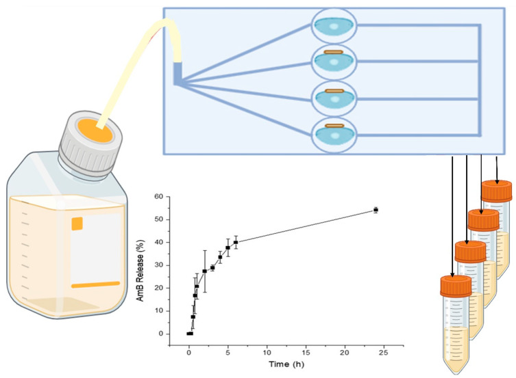
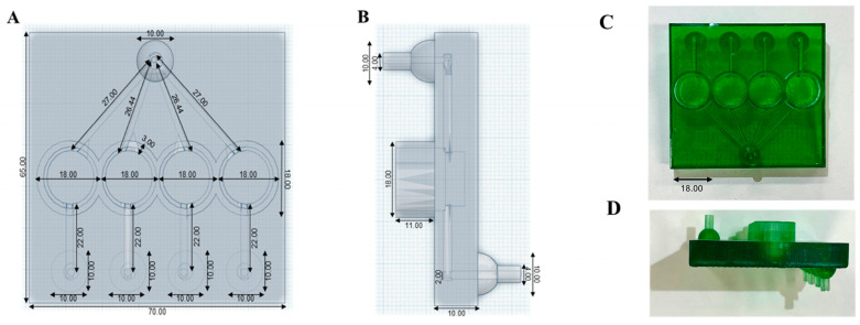
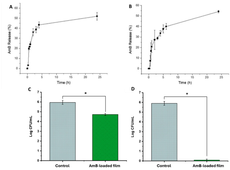
Fungal keratitis (FK), a severe eye infection that leads to vision impairment and blindness, poses a high risk to contact lens users, and Candida albicans remains the most common underpinning fungal pathogen in temperate climates. Patients are initially treated empirically (econazole 1% drops hourly for 24-48 h), and if there is no response, amphotericin B (AmB) 0.15% eye drops (extemporaneously manufactured to be stable for a week) are the gold-standard treatment. Here, we aim to develop a sustained-release AmB ocular film to treat FK with an enhanced corneal retention time. As there is a paucity of reliable in vitro models to evaluate ocular drug release and antifungal efficacy under flow, we developed a 3D-printed microfluidic device based on four chambers stacked in parallel, in which lenses previously inoculated with a C. albicans suspension were placed. Under the flow of a physiological fluid over 24 h, the release from the AmB-loaded film that was placed dry onto the surface of the wetted contact lenses was quantified, and their antifungal activity was assessed. AmB sodium deoxycholate micelle (dimeric form) was mixed with sodium alginate and hyaluronic acid (3:1 w/w) and cast into films (0.48 or 2.4%), which showed sustained release over 24 h and resulted in a 1.23-fold reduction and a 5.7-fold reduction in CFU/mL of C. albicans, respectively. This study demonstrates that the sustained delivery of dimeric AmB can be used for the treatment of FK and provides a facile in vitro microfluidic model for the development and testing of ophthalmic antimicrobial therapies.

Keywords: 3D printing; aggregation state; amphotericin B dimer; amphotericin b; keratitis; microfluidics; ocular films.


Conflict of interest statement

The authors declare no conflicts of interest.

Figures

Figure 1
Figure 1
Schematic representation of 3D-printed microfluidic chip to assess release from film under continuous flow.
Figure 2
Figure 2
Solid-state characterization analysis (top) (A): X-ray diffraction pattern: (a) AmB-loaded film, (b) lyophilized AmB dimer, (c) unprocessed hyaluronic acid, and (d) unprocessed sodium alginate. (B) Fourier-transform infrared (FTIR) spectra: (a) AmB-loaded film, (b) lyophilized AmB dimer, (c) unprocessed hyaluronic acid, and (d) unprocessed sodium alginate. Thermal characterization analysis (bottom): (C) differential scanning calorimetry (DSC) analysis and (D) thermogravimetric analysis (TGA) of AmB film (green line), AmB dimer (blue line), hyaluronic acid (red line), and sodium alginate (black line).
Figure 3
Figure 3
Mechanical properties of AmB-loaded films. (A) Adhesion to contact lens; (B) burst strength of AmB-loaded films. Shadow area in red indicates the standard deviation between measurements (n = 3).
Figure 4
Figure 4
Transmission electron microscopy (TEM) microphotographs. (A) AmB dimer; (B) AmB film stained with 1% phosphotungstic acid. Bar: 200 nm.
Figure 5
Figure 5
Design and dimensions of the 3D-printed microfluidic device. (A) Top view of the 3D-printed chip design. (B) Side view of the 3D-printed chip design (mm). (C) Top view of the 3D-printed ocular chip. (D) Side view of the 3D-printed ocular chip (mm). Dimensions are expressed in mm.
Figure 6
Figure 6
AmB release from (A) AmB film (0.48% w/w) and from (B) AmB film (2.4% w/w), and in vitro antifungal activity of the (C) AmB film (0.48% w/w) and (D) AmB film (2.4% w/w) versus control (blank untreated contact lenses). Statistical significant differences (p< 0.05) are depicted with *.

References

    1. Ting D.S.J., Galal M., Kulkarni B., Elalfy M.S., Lake D., Hamada S., Said D.G., Dua H.S. Clinical Characteristics and Outcomes of Fungal Keratitis in the United Kingdom 2011–2020: A 10-Year Study. J. Fungi. 2021;7:966. doi: 10.3390/jof7110966. - DOI - PMC - PubMed
    1. Lalitha P., Shapiro B.L., Srinivasan M., Prajna N.V., Acharya N.R., Fothergill A.W., Ruiz J., Chidambaram J.D., Maxey K.J., Hong K.C., et al. Antimicrobial susceptibility of Fusarium, Aspergillus, and other filamentous fungi isolated from keratitis. Arch. Ophthalmol. 2007;125:789–793. doi: 10.1001/archopht.125.6.789. - DOI - PubMed
    1. Hoffman J.J., Arunga S., Mohamed Ahmed A.H.A., Hu V.H., Burton M.J. Management of Filamentous Fungal Keratitis: A Pragmatic Approach. J. Fungi. 2022;8:1067. doi: 10.3390/jof8101067. - DOI - PMC - PubMed
    1. Watchaputi K., Jayasekara L., Ratanakhanokchai K., Soontorngun N. Inhibition of cell cycle-dependent hyphal and biofilm formation by a novel cytochalasin 19,20-epoxycytochalasin Q in Candida albicans. Sci. Rep. 2023;13:9724. doi: 10.1038/s41598-023-36191-4. - DOI - PMC - PubMed
    1. Yuste I., Luciano F.C., Anaya B.J., Sanz-Ruiz P., Ribed-Sanchez A., Gonzalez-Burgos E., Serrano D.R. Engineering 3D-Printed Advanced Healthcare Materials for Periprosthetic Joint Infections. Antibiotics. 2023;12:1229. doi: 10.3390/antibiotics12081229. - DOI - PMC - PubMed

LinkOut - more resources