Comparative Study
. 2024 Nov 10;22(11):509.
doi: 10.3390/md22110509.

Comparison of Microalgae Nannochloropsis oceanica and Chlorococcum amblystomatis Lipid Extracts Effects on UVA-Induced Changes in Human Skin Fibroblasts Proteome

Affiliations
Comparative Study

Comparison of Microalgae Nannochloropsis oceanica and Chlorococcum amblystomatis Lipid Extracts Effects on UVA-Induced Changes in Human Skin Fibroblasts Proteome

Sinemyiz Atalay Ekiner et al. Mar Drugs. .

Abstract

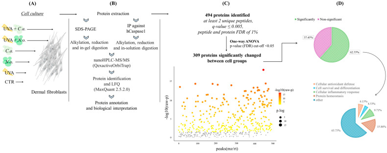
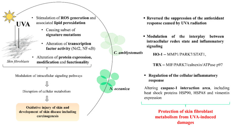
Lipid extracts from the microalgae Nannochloropsis oceanica and Chlorococcum amblystomatis have great potential to prevent ultraviolet A (UVA)-induced metabolic disorders. Therefore, the aim of this study has been to analyze their cytoprotective effect, focused on maintaining intracellular redox balance and inflammation in UVA-irradiated skin fibroblasts, at the proteome level. The above lipid extracts reversed the suppression of the antioxidant response caused by UVA radiation, which was more visible in the case of C. amblystomatis. Modulations of interactions between heme oxygenase-1 and matrix metalloproteinase 1/Parkinson's disease protein 7/transcript1-α/β, as well as thioredoxin and migration inhibitory factor/Parkinson's disease protein 7/calnexin/ATPase p97, created key molecular signaling underlying their cytoprotective actions. Moreover, they reduced pro-inflammatory processes in the control group but they also showed the potential to regulate the cellular inflammatory response by changing inflammasome signaling associated with the changes in the caspase-1 interaction area, including heat shock proteins HSP90, HSPA8, and vimentin. Therefore, lipid extracts from N. oceanica and C. amblystomatis protect skin fibroblast metabolism from UVA-induced damage by restoring the redox balance and regulating inflammatory signaling pathways. Thus, those extracts have proven to have great potential to be used in cosmetic or cosmeceutical products to protect the skin against the effects of solar radiation. However, the possibility of their use requires the evaluation of their effects at the skin level in in vivo and clinical studies.

Keywords: Chlorococcum amblystomatis; Nannochloropsis oceanica; UVA radiation; cytoprotective effect; fibroblast; inflammation; lipid extracts; oxidative stress; proteomics.


Conflict of interest statement

The authors declare no conflicts of interest.

Figures

Figure 1
Figure 1
Summary of the experimental design presenting the experimental cell groups [preparation of the CTR, N.o., C.a., UVA, UVA + N.o., and UVA + C.a. cell groups, as explained in Table 1] (A), proteomic sample preparation including SDS-PAGE or IP (immunoprecipitation) and MS/MS proteomic analysis (B), as well as the obtained complete proteomic data with the main statistical thresholds used (C) and a general assessment of the obtained protein groups highlighting their biological functions (cellular antioxidant defense/cell survival and differentiation/cellular inflammatory response/protein homeostasis), and the percentage of identified protein numbers (for each protein group) compared to the number of total identified proteins (D). “Significant” is used for the proteins whose intensities are statistically significantly changed between experimental cell groups according to the one-way ANOVA. “Non-significant” is used for the proteins whose intensities are not changed statistically significantly between cell groups according to the one-way ANOVA. (hCaspase1, human caspase-1; LFQ, label-free quantification; FDR, false discovery rate; nanoHPLC, nano-high-performance liquid chromatography; MS, mass spectrometry; QexactiveOrbiTrap, Q Exactive HF mass spectrometer.)
Figure 2
Figure 2
Principal component analysis (PCA) of the skin fibroblasts’ proteome from the experimental cell groups CTR, N.o., C.a., UVA, UVA + N.o., and UVA + C.a. showing an obvious clustering of CTR and UVA samples in distinct groups. (CTR, control cells cultured in the standard growing medium; N.o., cells cultured in the standard growing medium also containing lipid extracts from N. oceanica (2 µg/mL) for 24 h; C.a., cells cultured in the standard growing medium containing lipid extracts from C. amblystomatis (2 µg/mL) for 24 h; UVA, cells exposed to UVA (365 nm) at a dose of 13 J/cm3 and then incubated in the standard growing medium for 24 h; UVA + N.o., cells exposed to UVA (365 nm) at a dose of 13 J/cm3 and then incubated in the standard growing medium containing lipid extracts from N. oceanica (2 µg/mL) for 24 h; UVA + C.a., cells exposed to UVA (365 nm) at a dose of 13 J/cm3 and then incubated in the standard growing medium containing lipid extracts from C. amblystomatis (2 µg/mL) for 24 h; PC1/2/3, principal component 1/2/3.)
Figure 3
Figure 3
Volcano plots comparing the effects of N. oceanica and C. amblystomatis lipid extracts on the proteome of skin fibroblasts from the experimental cell groups (CTR, control cells cultured in the standard growing medium; N.o., cells cultured in the standard growing medium also containing lipid extracts from N. oceanica (2 µg/mL) for 24 h; C.a., cells cultured in the standard growing medium containing lipid extracts from C. amblystomatis (2 µg/mL) for 24 h; UVA, cells exposed to UVA (365 nm) at a dose of 13 J/cm3 and then incubated in the standard growing medium for 24 h; UVA + N.o., cells exposed to UVA (365 nm) at a dose of 13 J/cm3 and then incubated in the standard growing medium containing lipid extracts from N. oceanica (2 µg/mL) for 24 h; UVA + C.a., cells exposed to UVA (365 nm) at a dose of 13 J/cm3 and then incubated in the standard growing medium containing lipid extracts from C. amblystomatis (2 µg/mL) for 24 h). [Significant features (in blue and red as seen in figure—fold change) had p < 0.05].
Figure 4
Figure 4
Heatmap, clustering of the top 25 proteins with significantly changed expressions in the skin fibroblasts of the following cell groups: control group (CTR), N. oceanica or C. amblystomatis lipid extract-treated group (N.o./C.a.), UVA-irradiated cell group (UVA), and cell group treated with N. oceanica or C. amblystomatis lipid extracts following UVA irradiation (UVA + N.o./UVA + C.a.), as explained in Table 1. While the protein functions of the mentioned proteins were included, the proteins involved in the regulation of inflammation or protein homeostasis were also indicated.
Figure 5
Figure 5
Profile plot generated by the Perseus software platform (version 2.0.11.0), using the average intensities (log-transformed and normalized by median) of proteins with cytoprotective activity against oxidative stress whose levels were significantly changed between cell groups (CTR, control cells cultured in the standard growing medium; N.o., cells cultured in the standard growing medium also containing lipid extracts from N. oceanica (2 µg/mL) for 24 h; C.a., cells cultured in the standard growing medium containing lipid extracts from C. amblystomatis (2 µg/mL) for 24 h; UVA, cells exposed to UVA (365 nm) at a dose of 13 J/cm3 and then incubated in the standard growing medium for 24 h; UVA + N.o., cells exposed to UVA (365 nm) at a dose of 13 J/cm3 and then incubated in the standard growing medium containing lipid extracts from N. oceanica (2 µg/mL) for 24 h; UVA + C.a., cells exposed to UVA (365 nm) at a dose of 13 J/cm3 and then incubated in the standard growing medium containing lipid extracts from C. amblystomatis (2 µg/mL) for 24 h). Only the proteins showing at least 3-fold changes (log2FC) were highlighted with pink, blue, green, and yellow colors (different than grey color). Except for thioredoxin, N. oceanica or C. amblystomatis lipid extracts were able to reverse UVA-disrupted expression of aldo-keto reductase family member A1, peroxiredoxin-5, mitochondrial, and heme oxygenase-1 towards their CTR-level expressions. Statistics for the indicated proteins (mean values ± SD of three independent samples and statistically significant differences for p ≤ 0.05) are presented in Supplementary Figure S6.
Figure 6
Figure 6
Profile plot generated by the Perseus software platform (version 2.0.11.0), using the average intensities (log-transformed and normalized by median) of proteins participating in the regulation of cellular inflammatory response whose levels were significantly changed between cell groups (CTR, control cells cultured in the standard growing medium; N.o., cells cultured in the standard growing medium also containing lipid extracts from N. oceanica (2 µg/mL) for 24 h; C.a., cells cultured in the standard growing medium containing lipid extracts from C. amblystomatis (2 µg/mL) for 24 h; UVA, cells exposed to UVA (365 nm) at a dose of 13 J/cm3 and then incubated in the standard growing medium for 24 h; UVA + N.o., cells exposed to UVA (365 nm) at a dose of 13 J/cm3 and then incubated in the standard growing medium containing lipid extracts from N. oceanica (2 µg/mL) for 24 h; UVA + C.a., cells exposed to UVA (365 nm) at a dose of 13 J/cm3 and then incubated in the standard growing medium containing lipid extracts from C. amblystomatis (2 µg/mL) for 24 h). Only the proteins showing at least 3-fold changes (log2FC) were highlighted with different colors (different than grey). Statistics for the indicated proteins (mean values ± SD of three independent samples and statistically significant differences for p ≤ 0.05) are presented in Supplementary Figure S7.
Figure 7
Figure 7
N. oceanica and C. amblystomatis lipid extracts restore the UVA-impaired control expression of antioxidant proteins and proteins involving inflammatory signaling in skin fibroblasts (ultraviolet A, UVA; reactive oxygen species, ROS; nuclear factor erythroid 2-related factor 2, Nrf2; nuclear factor kappa, NF-κB; heme oxygenase-1, HO-1; aldo-keto reductase family member 1 A1, AKR1A1; thioredoxin, Trx; matrix metalloproteinase-1, MMP1; Parkinson’s disease protein 7, PARK7; signal transducer and activator of transcription1-alpha/beta, STAT1; macrophage migration inhibitory factor, MIF).

References

    1. D’Orazio J., Jarrett S., Amaro-Ortiz A., Scott T. UV Radiation and the Skin. Int. J. Mol. Sci. 2013;14:12222–12248. doi: 10.3390/ijms140612222. - DOI - PMC - PubMed
    1. Byrd A.L., Belkaid Y., Segre J.A. The human skin microbiome. Nat. Rev. Microbiol. 2018;16:143–155. doi: 10.1038/nrmicro.2017.157. - DOI - PubMed
    1. Kong J., Hu X.-M., Cai W.-W., Wang Y.-M., Chi C.-F., Wang B. Bioactive Peptides from Skipjack Tuna Cardiac Arterial Bulbs (II): Protective Function on UVB-Irradiated HaCaT Cells through Antioxidant and Anti-Apoptotic Mechanisms. Mar. Drugs. 2023;21:105. doi: 10.3390/md21020105. - DOI - PMC - PubMed
    1. Zhao H.-C., Xiao T., Chen Y.-J. Ultraviolet Induced Skin Inflammation. Int. J. Dermatol. Venereol. 2021;4:229–235. doi: 10.1097/JD9.0000000000000144. - DOI
    1. Pfeifer G.P. Mechanisms of UV-induced mutations and skin cancer. Genome Instab Dis. 2020;1:99–113. doi: 10.1007/s42764-020-00009-8. - DOI - PMC - PubMed

Publication types

LinkOut - more resources