. 2024 Oct 25;12(11):1213.
doi: 10.3390/vaccines12111213.

B and T Cell Bi-Cistronic Multiepitopic Vaccine Induces Broad Immunogenicity and Provides Protection Against SARS-CoV-2

Affiliations

B and T Cell Bi-Cistronic Multiepitopic Vaccine Induces Broad Immunogenicity and Provides Protection Against SARS-CoV-2

Beatriz Perdiguero et al. Vaccines (Basel). .

Abstract

Background: The COVID-19 pandemic, caused by SARS-CoV-2, has highlighted the need for vaccines targeting both neutralizing antibodies (NAbs) and long-lasting cross-reactive T cells covering multiple viral proteins to provide broad and durable protection against emerging variants.

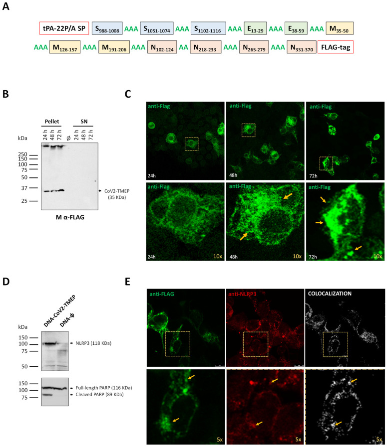
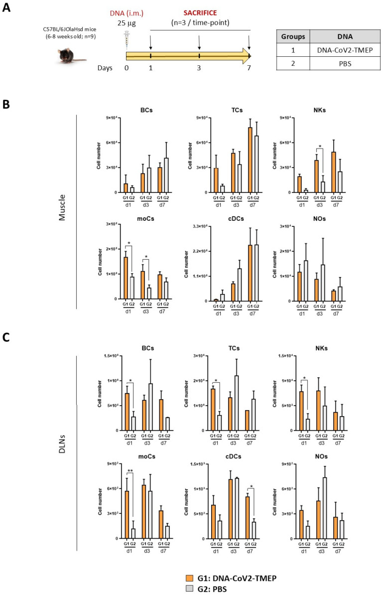
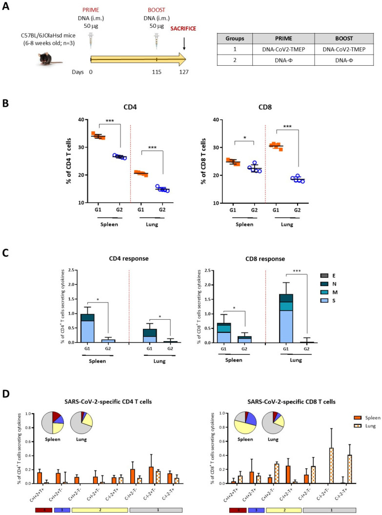
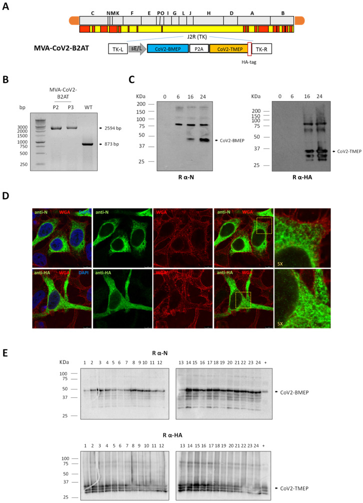
Methods: To address this, here we developed two vaccine candidates, namely (i) DNA-CoV2-TMEP, expressing the multiepitopic CoV2-TMEP protein containing immunodominant and conserved T cell regions from SARS-CoV-2 structural proteins, and (ii) MVA-CoV2-B2AT, encoding a bi-cistronic multiepitopic construct that combines conserved B and T cell overlapping regions from SARS-CoV-2 structural proteins.

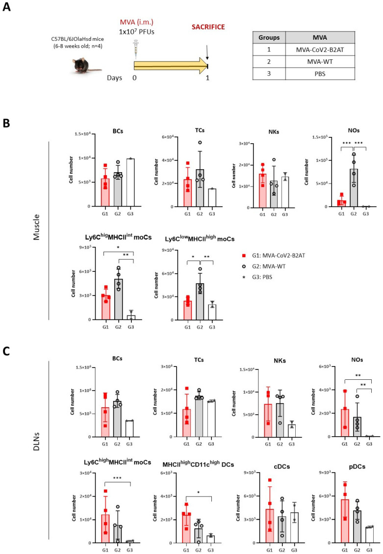
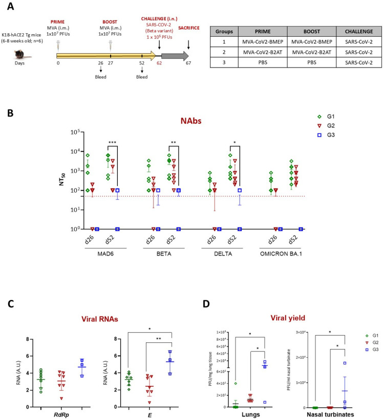
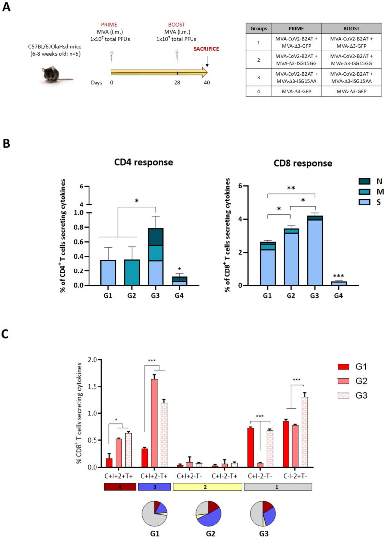
Results: Both candidates were assessed in vitro and in vivo demonstrating their ability to induce robust immune responses. In C57BL/6 mice, DNA-CoV2-TMEP enhanced the recruitment of innate immune cells and stimulated SARS-CoV-2-specific polyfunctional T cells targeting multiple viral proteins. MVA-CoV2-B2AT elicited NAbs against various SARS-CoV-2 variants of concern (VoCs) and reduced viral replication and viral yields against the Beta variant in susceptible K18-hACE2 mice. The combination of MVA-CoV2-B2AT with a mutated ISG15 form as an adjuvant further increased the magnitude, breadth and polyfunctional profile of the response.

Conclusion: These findings underscore the potential of these multiepitopic proteins when expressed from DNA or MVA vectors to provide protection against SARS-CoV-2 and its variants, supporting their further development as next-generation COVID-19 vaccines.

Keywords: DNA and MVA vectors; ISG15 adjuvant; SARS-CoV-2; cellular response; efficacy in mice; immunogenicity; innate response; multiepitopic protein; vaccine.


Conflict of interest statement

The authors declare no conflicts of interest.

Figures

Figure 1
Figure 1
In vitro characterization of a DNA vector expressing the multiepitopic CoV2-TMEP protein. (A) Schematic representation of the chimeric CoV2-TMEP multiepitopic vaccine targeting T cells. (B) Time-course expression of CoV2-TMEP protein. HEK-293T cells were transfected as detailed in the Materials and Methods section and CoV2-TMEP expression in pellets and supernatants (SN) was analyzed by Western blotting using the mouse monoclonal anti-FLAG antibody. (C) Subcellular localization of CoV2-TMEP protein. HeLa cells were transfected and processed for immunofluorescence microscopy as indicated in the Materials and Methods section. Green staining: mouse monoclonal anti-FLAG antibody followed by anti-mouse Alexa Fluor 488 antibody. Yellow arrows: CoV2-TMEP forming aggregates in magnified images (10×). (D) Analysis of NLRP3 induction and PARP cleavage. HEK-293T cells were transfected as described in the Materials and Methods section and the expression of NLRP3 (118 KDa; upper panel) and PARP (full-length: 116 KDa; cleaved: 89 KDa; lower panel) was analyzed by Western blotting using specific antibodies. (E) Subcellular colocalization of CoV2-TMEP protein with inflammasome. HeLa cells were transfected and processed for confocal microscopy as detailed in the Materials and Methods section. A mouse monoclonal anti-FLAG antibody and a rabbit polyclonal anti-NLRP3/NALP3 antibody followed by the corresponding secondary antibodies conjugated with Alexa Fluor 488 (green staining; FLAG detection) or Alexa Fluor 594 (red staining; NLRP3 detection) were used. Colocalization signal is indicated in white. Yellow arrows: Discrete colocalization signals in magnified images (5×). One representative optical section from three independent experiments is shown.
Figure 2
Figure 2
Innate immune response elicited in muscle and DLNs from mice immunized with a DNA vector expressing the polyvalent multiepitopic CoV2-TMEP protein. (A) Immunization schedule. Mice were inoculated with DNA-CoV2-TMEP or PBS by intramuscular (i.m.) route. At the indicated days post-inoculation, total muscle from the site of inoculation and DLNs were harvested and processed as described in the Materials and Methods section. (B,C) Immune cell populations present in muscle (B) and DLNs (C) determined by flow cytometry. Data are shown as mean and SD. BCs: B cells; TCs: T cells; NKs: natural killer cells; moCs: monocyte-derived cells; cDCs: conventional dendritic cells; NOs: neutrophils. *, p < 0.05; **, p < 0.005.
Figure 3
Figure 3
SARS-CoV-2-specific T cell adaptive immune responses elicited by DNA-CoV2-TMEP in C57BL/6 mice when administered in homologous regimen. (A) Immunization schedule. Mice were inoculated with DNA-CoV2-TMEP or DNA-ϕ by i.m. route at days 0 and 115. At 12 days post-boost, animals were sacrificed, and spleens and lungs were harvested and processed for ICS assay, as described in the Materials and Methods section. (B) Magnitude of the total CD4 (left panel) or CD8 (right panel) T cells in the spleens and lungs of immunized mice. Each colored form represents the value obtained for each stimulus (S, M, N, E and RPMI). The 95% confidence interval (CI) is represented. (C) Magnitude of the total SARS-CoV-2-specific CD4 (left) or CD8 (right) T cells at 12 days post-boost after the stimulation of lymphocytes derived from spleen or lung with SARS-CoV-2 peptide pools. The total value of each group represents the sum of the percentages of SARS-CoV-2-specific CD4 or CD8 T cells expressing CD107a and/or producing IFN-γ and/or IL-2 and/or TNF-α against SARS-CoV-2 peptide pools. Data are background-subtracted. The 95% CI is represented. (D) Polyfunctional profile of the SARS-CoV-2-specific CD4 (left) or CD8 (right) T cells in DNA-CoV2-TEMP-immunized mice in spleen and lung. The positive combinations of the responses are indicated on the x axis, while the percentages of the functionally different cell populations within the total CD4 or CD8 T cells are represented on the y axis. Specific responses are grouped and color-coded based on the number of functions. All data are background-subtracted. The 95% CI is shown. C: CD107a; I: IFN-γ; 2: IL-2; T: TNF-α. *, p < 0.05; ***, p < 0.001.
Figure 4
Figure 4
In vitro characterization of an MVA-based recombinant virus expressing the multiepitopic CoV2-BMEP-P2A-TMEP protein. (A) Scheme of the thymidine kinase (TK) locus of MVA-CoV2-B2AT recombinant virus. (B) Confirmation of CoV2-BMEP-P2A-TMEP gene insertion. DNA extracted from DF-1 cells infected with MVA-WT or MVA-CoV2-B2AT (P2 and P3 viral preparations) viruses as described in the Materials and Methods section was used as template for the analysis of the TK locus by PCR using primers TK-L and TK-R spanning TK flanking regions. (C) Time-course expression of CoV2-BMEP-P2A-TMEP protein. DF-1 cells infected with MVA-CoV2-B2AT as detailed in the Materials and Methods section were harvested at the indicated times post-infection and analyzed by Western blotting using a rabbit polyclonal anti-SARS-CoV-2 N antibody (left; CoV2-BMEP) or a rabbit polyclonal anti-SARS-CoV-2 HA antibody (right; CoV2-TMEP). (D) Subcellular localization of CoV2-BMEP-P2A-TMEP protein. HeLa cells infected with MVA-CoV2-B2AT for 16 h were harvested and processed for immunofluorescence microscopy as described in the Materials and Methods section. Red staining: WGA-Alexa Fluor 555; green staining: rabbit polyclonal anti-N antibody (upper panels) or rabbit polyclonal anti-HA antibody (lower panels) followed by the corresponding secondary antibody conjugated with Alexa Fluor 488; blue staining: DAPI. (E) Stability of the CoV2-BMEP-P2A-TMEP protein. The expression of CoV2-BMEP-P2A-TMEP protein in 24 individual plaques from MVA-CoV2-B2AT stability passage 7 was analyzed by Western blotting using a rabbit polyclonal anti-N antibody (upper) or a rabbit polyclonal anti-HA antibody (lower). Lane (+): MVA-CoV2-B2AT P2 stock.
Figure 5
Figure 5
Innate immune response elicited in muscle and DLNs from mice immunized with an MVA vector expressing the multiepitopic CoV2-BMEP-P2A-TMEP protein. (A) Immunization schedule. Mice were immunized with MVA-WT or MVA-CoV2-B2AT or PBS-treated by i.m. route. At day 1 post-inoculation, total muscle from the site of inoculation and DLNs were excised and processed as described in the Materials and Methods section. (B,C) Immune cell populations present in muscle (B) and DLNs (C) determined by flow cytometry. Data are shown as colored forms for each animal with mean and SD. BCs: B cells; TCs: T cells; NKs: natural killer cells; NOs: neutrophils; moCs: monocyte-derived cells; DCs: dendritic cells; cDCs: conventional dendritic cells; pDCs: plasmacytoid dendritic cells. *, p < 0.05; **, p < 0.005; ***, p < 0.001.
Figure 6
Figure 6
Efficacy study in K18-hACE2 Tg mice. (A) Immunization schedule. K18-hACE2 Tg mice were immunized by i.m. route with the indicated viruses or with PBS at days 0 and 27 and challenged 5 weeks later (d62) by intranasal (i.n.) route with SARS-CoV-2 virus (Beta variant). At 5 days post-challenge, mice were sacrificed, and lung, nasal turbinate and serum samples were harvested and processed as described in the Materials and Methods section. (B) SARS-CoV-2 NAbs (NT50) in serum from immunized mice against SARS-CoV-2 MAD6 and Beta, Delta and Omicron BA.1 VoCs at d26 and d52 determined by MNT assay. Data are shown as forms for each animal with mean and SD. Red dashed line: lower limit of detection (LLD) of the assay. (C) Virus replication in lung samples. Genomic (RdRp) and subgenomic (E) SARS-CoV-2 RNAs were detected by RT-qPCR post-challenge. RNA levels [in arbitrary units (A.U.)] for each animal are represented as forms with mean and SD from duplicates; relative values are referred to as a naïve lung sample. (D) SARS-CoV-2 infectious virus in lungs (left) and nasal turbinates (right). Data are shown as forms for each animal with mean (PFUs/mg of lung tissue or PFUs/mL of nasal turbinate tissue) and SD from duplicates. *, p < 0.05; **, p < 0.005; ***, p < 0.001.
Figure 7
Figure 7
SARS-CoV-2-specific T cell adaptive immune responses elicited by MVA-CoV2-B2AT in C57BL/6 mice when administered in homologous regimen in the presence or absence of ISG15 as adjuvant. (A) Immunization schedule. Mice were inoculated with the indicated vector combinations by i.m. route at days 0 and 28. At 12 days post-boost, animals were sacrificed and spleens were harvested and processed for ICS assay, as described in the Materials and Methods section. (B) Magnitude of the total SARS-CoV-2-specific CD4 (left) or CD8 (right) T cells at 12 days post-boost after the stimulation of splenocytes with SARS-CoV-2 peptide pools. The total value of each group represents the sum of the percentages of SARS-CoV-2-specific CD4 or CD8 T cells expressing CD107a and/or producing IFN-γ and/or IL-2 and/or TNF-α against SARS-CoV-2 peptide pools. Data were background-subtracted. The 95% CI is represented. (C) Polyfunctional profile of the SARS-CoV-2-specific CD8 T cells in immunized mice. The positive combinations of the responses are indicated on the x axis, while the percentages of the functionally different cell populations within the total CD8 T cells are represented on the y axis. Specific responses are grouped and color-coded based on the number of functions. All data were background-subtracted. The 95% CI is shown. C: CD107a; I: IFN-γ; 2: IL-2; T: TNF-α. *, p < 0.05; **, p < 0.005; ***, p < 0.001.

References

    1. Senevirathne T.H., Wekking D., Swain J.W.R., Solinas C., De Silva P. COVID-19: From emerging variants to vaccination. Cytokine Growth Factor Rev. 2024;76:127–141. doi: 10.1016/j.cytogfr.2023.11.005. - DOI - PubMed
    1. Cohen K.W., Linderman S.L., Moodie Z., Czartoski J., Lai L., Mantus G., Norwood C., Nyhoff L.E., Edara V.V., Floyd K., et al. Longitudinal analysis shows durable and broad immune memory after SARS-CoV-2 infection with persisting antibody responses and memory B and T cells. Cell Rep. Med. 2021;2:100354. doi: 10.1016/j.xcrm.2021.100354. - DOI - PMC - PubMed
    1. Sette A., Crotty S. Adaptive immunity to SARS-CoV-2 and COVID-19. Cell. 2021;184:861–880. doi: 10.1016/j.cell.2021.01.007. - DOI - PMC - PubMed
    1. Heitmann J.S., Bilich T., Tandler C., Nelde A., Maringer Y., Marconato M., Reusch J., Jager S., Denk M., Richter M., et al. A COVID-19 peptide vaccine for the induction of SARS-CoV-2 T cell immunity. Nature. 2022;601:617–622. doi: 10.1038/s41586-021-04232-5. - DOI - PMC - PubMed
    1. Meyers L.M., Gutierrez A.H., Boyle C.M., Terry F., McGonnigal B.G., Salazar A., Princiotta M.F., Martin W.D., De Groot A.S., Moise L. Highly conserved, non-human-like, and cross-reactive SARS-CoV-2 T cell epitopes for COVID-19 vaccine design and validation. NPJ Vaccines. 2021;6:71. doi: 10.1038/s41541-021-00331-6. - DOI - PMC - PubMed

LinkOut - more resources