. 2024 Nov 26;17(864):eadp1375.
doi: 10.1126/scisignal.adp1375. Epub 2024 Nov 26.

Coordination between the eIF2 kinase GCN2 and p53 signaling supports purine metabolism and the progression of prostate cancer

Affiliations

Coordination between the eIF2 kinase GCN2 and p53 signaling supports purine metabolism and the progression of prostate cancer

Ricardo A Cordova et al. Sci Signal. .

Abstract

Cancers invoke various pathways to mitigate external and internal stresses to continue their growth and progression. We previously reported that the eIF2 kinase GCN2 and the integrated stress response are constitutively active in prostate cancer (PCa) and are required to maintain amino acid homeostasis needed to fuel tumor growth. However, although loss of GCN2 function reduces intracellular amino acid availability and PCa growth, there is no appreciable cell death. Here, we discovered that the loss of GCN2 in PCa induces prosenescent p53 signaling. This p53 activation occurred through GCN2 inhibition-dependent reductions in purine nucleotides that impaired ribosome biogenesis and, consequently, induced the impaired ribosome biogenesis checkpoint. p53 signaling induced cell cycle arrest and senescence that promoted the survival of GCN2-deficient PCa cells. Depletion of GCN2 combined with loss of p53 or pharmacological inhibition of de novo purine biosynthesis reduced proliferation and enhanced cell death in PCa cell lines, organoids, and xenograft models. Our findings highlight the coordinated interplay between GCN2 and p53 regulation during nutrient stress and provide insight into how they could be targeted in developing new therapeutic strategies for PCa.


Conflict of interest statement

RCW is a member of the advisory board of HiberCell, Inc. KAS is a consultant for HiberCell, Inc. and Aclaris Therapeutics and receives research support from HiberCell, Inc. TGA consults for HiberCell, Inc.

Figures
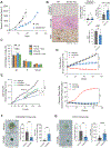
Figure 1.
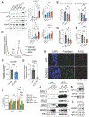
Figure 1.. Loss of GCN2 leads to a slow growth phenotype in PCa that is characterized by increased senescence and minimal cell death.
(A) Tumor volumes on the indicated days in male NSG mice bearing subcutaneous xenografts of WT or GCN2 KO 22Rv1 cells (N = 6 mice per group). Statistical significance was determined using a two-way repeated measures analysis of variance (ANOVA) with Sidak’s multiple comparisons. Error bars indicate the standard error of the mean (SEM). (B) Representative images and analysis for cleaved caspase 3, β-galactosidase (SA-β-gal), and Ki-67 immunohistochemistry (IHC) staining in WT and GCN2 KO 22Rv1 tumors described in (A). Quantification for each stain is displayed as percent positive cells (N = 5 tumor sections per group, each from an independent mouse). Statistical significance was determined using an unpaired two-tailed t-test. (C) Cell cycle analyses of LNCaP cells treated with vehicle, GCN2iB (5 μM), vehicle and EAA, or GCN2iB (5 μM) and EAA for 48 hours. Statistical significance was determined using a two-way ANOVA with Tukey’s multiple comparisons (N = 3 biological replicates). Error bars indicate the standard deviation (SD). (D) LNCaP cells were treated with vehicle, GCN2iB (5 μM), or thapsigargin (1 μM) as a positive control (+ C). Cell proliferation (top) and cell death (bottom; by cytotox red stain) were measured every 4 hours using an IncuCyte System. Statistical significance was determined using a two-way ANOVA with Sidak’s multiple comparisons (N = 5 biological replicates). Error bars indicate the SD. (E) LNCaP cells were treated with vehicle or GCN2iB (2 μM). After 64 hours, treatment media was removed, cells were washed and cultured with media supplemented with GCN2iB (5 μM), GCN2iB (5 μM) + EAA, or vehicle as indicated. Cell growth was measured using an IncuCyte system and statistical significance was determined using a two-way ANOVA with Tukey’s multiple comparisons (N = 5 biological replicates). (F and G) Representative images of (F) TM00298 and (G) DKO (Pten−/− Rb1−/−) PCa organoids treated with GCN2iB for 10 or 5 days, respectively, and stained for SA-β-gal. Bar graphs indicate the average organoid area (mm2) and percent positive organoid area stained for SA-β-gal (N ≥ 6 organoids per group). Statistical significance was determined using an unpaired two-tailed t-test. Error bars indicate SD. For all panels, ns, non-significant, *p ≤ 0.05, **p ≤ 0.001, ***p ≤ 0.001, and ****p ≤ 0.0001 by the test indicated.
Figure 2.
Figure 2.. Inhibition of GCN2 and the ISR induce p53 signaling in PCa cells.
(A) Volcano plot indicating genes altered in expression in LNCaP cells treated with GCN2iB using an RNA-seq dataset that we previously reported (10). The fold change for gene transcripts with adjusted p-value (−log10) comparing LNCaP cells treated with GCN2iB (2 μM) versus vehicle control for 24 hours (increased and decreased set as ≥ 2-fold, fdr ≤ 0.05). ISR-dependent genes are highlighted in green, and p53 target genes are highlighted in red. (B) Heatmap illustrating the top canonical pathways as determined using Ingenuity Pathway Analysis (IPA) for LNCaP cells treated with GCN2iB (2 μM) for 6 or 24 hours compared to vehicle (data file S1). The scale bar indicates the Z score. (C) Heatmap displaying significantly expressed genes associated with indicated canonical pathways from panel (A). The scale bar indicates fold change in gene expression. (D) K-means clustering (k = 2; N = 1000 transcripts) from RNA-seq transcriptome data of LNCaP cells treated with vehicle or GCN2iB (2 μM) for 6 or 24 hours (data file S1). The number of genes per cluster is indicated, and the scale bar represents normalized read counts. Bar graphs show the top enriched gene sets and their respective significance (−log10 p-value) for each gene cluster (data file S1). (E) Lysates were prepared from LNCaP cells treated with vehicle or GCN2iB (5 μM) for 24 or 48 hours, and immunoblot analyses were carried out using antibodies that recognize phosphorylated or total GCN2, ATF4, p53, p21, MDM2, or actin. ATF4 is indicated by the arrow. Molecular weight in kilodaltons (kDa) is marked. Levels of the indicated proteins normalized to controls are shown in the bar graphs. Statistical significance was determined using a one-way ANOVA with Tukey’s multiple comparisons (N = 4 biological replicates). Error bars indicate SD. (F) CDKN1A (p21), MDM2, GADD45A, and ASNS mRNA were measured by qRT-PCR in LNCaP cells treated with vehicle or GCN2iB (5 μM) for 24 or 48 hours. Statistical significance was determined using a one-way ANOVA with Tukey’s multiple comparisons (N = 4 biological replicates). Error bars indicate SD. For all panels, *p ≤ 0.05, **p ≤ 0.001, ***p ≤ 0.001, and ****p ≤ 0.0001 by the test indicated.
Figure 3.
Figure 3.. GCN2 and p53 coordinate amino acid homeostasis and metabolism in PCa.
(A) Venn diagram displaying differentially expressed genes from RNA-seq transcriptome analysis of LNCaP WT or LNCaP p53 KO cells treated with vehicle or GCN2iB (5 μM) for 24 hours (N = 4 samples per group; data file S2). (B and C) IPA Upstream Regulator analysis from the transcriptome analyses. ATF4 (B) and p53 (C) are shown as top upstream regulators, and the scale bar indicates the z score (data file S2). (D) Heatmaps featuring ATF4 (middle) and p53 (right) dependent genes. The scale bar shows log2 fold change in gene expression (data file S2). (E) K-means clustering (k = 4; N = 1800 transcripts; data file S2) of gene expression data presented in (A). The number of genes per cluster is indicated, and the scale bar represents normalized read counts. Bar graphs show the top enriched gene sets and their respective significance (-log10 p-value) for each gene cluster (data file S2).
Figure 4.
Figure 4.. Induction of p53 following GCN2 inhibition in PCa cells results in a G1 cell cycle arrest, increased senescence, and promotes cell survival.
(A) Lysates were prepared from LNCaP WT or LNCaP p53 KO cells treated with GCN2iB (5 μM) or vehicle for 48 hours and analyzed by immunoblot using antibodies that recognize the indicated proteins. Levels of the indicated proteins normalized to control are shown in the bar graph. Statistical significance was determined using a one-way ANOVA with Tukey’s multiple comparisons (N = 3 biological replicates). Error bars indicate SD. (B) Cell cycle analysis of LNCaP WT or LNCaP p53 KO cells treated with GCN2iB (5 μM) or vehicle for 48 hours. Statistical significance was determined using a two-way ANOVA with Tukey’s multiple comparisons (N = 3 biological replicates). Error bars indicate SD. (C) Representative images of DKO (Pten−/− Rb1−/−) and TKO (Pten−/− Rb1−/− TP53−/−) organoids treated with GCN2iB (5 μM) for 5 days and stained with Cytotox Red Dye and Hoechst 33342. Bar graphs indicate the average organoid area (mm2) and percent organoid area staining positive with Cytotox Red. Statistical significance was determined using a two-way ANOVA with Tukey’s multiple comparisons (N ≥12 organoids per group). Error bars indicate SD. (D) DKO and TKO organoids were treated as described in (C) and were stained and analyzed for SA-β-gal. Statistical significance was determined using a two-way ANOVA with Tukey’s multiple comparisons (N ≥ 15 organoids per group). Error bars indicate SD. (E) LNCaP cells were treated with vehicle, GCN2iB (5 μM), vehicle and EAA, or GCN2iB (5 μM) and EAA for 48 hours. Lysates were prepared and analyzed by immunoblot analysis using antibodies for the indicated proteins. The levels of the indicated proteins normalized to control are shown in the bar graphs. Statistical significance was determined using a one-way ANOVA with Tukey’s multiple comparisons (N = 3 biological replicates). Error bars indicate SD. (F) LNCaP cells were transfected with two different siRNAs targeting p53 or a scramble siRNA control. Cell lysates were prepared and analyzed by immunoblotting for the indicated proteins, quantified as. normalized to control. Statistical significance was determined using a one-way ANOVA with Tukey’s multiple comparisons (N = 3 biological replicates). Error bars indicate SD. (G) Levels of amino acids in LNCaP cells transfected with one of two different siRNAs targeting p53 or a scrambled siRNA control. Bar graphs show the levels of high abundance (left) and low abundance (right) amino acids. Statistical significance was determined using a two-way ANOVA with Tukey’s multiple comparisons (N = 4 biological replicates). Error bars indicate SD. For all panels; *p ≤ 0.05, **p ≤ 0.001, ***p ≤ 0.001, and ****p ≤ 0.0001 by the test indicated.
Figure 5.
Figure 5.. GCN2 inhibition in PCa cells depletes purines leading to p53 activation.
(A) Untargeted metabolomics of LNCaP cells treated with GCN2iB (2 μM) or vehicle for 8 hours (data file S4). Volcano plot illustrating log2 fold change in metabolite levels with adjusted p-value (−log10) comparing LNCaP cells treated with GCN2iB vs vehicle. Increased metabolites are highlighted in red, and decreased metabolites are shown in green (fdr ≤ 0.05). (N = 3 biological replicates). (B) Altered metabolites from untargeted metabolomics from (A) were analyzed using MetaboAnalyst to test for enriched pathways using the KEGG database (data file S4). Bubble plots indicate enriched metabolic pathways in GCN2iB-treated LNCaP cells. (C) LNCaP cells were treated with vehicle, GCN2iB (5 μM), vehicle + nucleobase (NB) mix (50 μM of each nucleobase), or GCN2iB (5 μM) + NB mix (50 μM of each nucleobase), and cell growth was measured for up to 6 days. Statistical significance was determined using a two-way ANOVA with Tukey’s multiple comparisons (N = 5 biological replicates). Error bars indicate SD. (D) LNCaP cells were treated with vehicle, GCN2iB (5 μM), or GCN2iB (5 μM) combined with the indicated individual nucleobases (50 μM) or D-ribose (5 mM), and cell growth was assessed after 2 days. Statistical significance was determined using a one-way ANOVA with Tukey’s multiple comparisons (N = 3 biological replicates). Error bars indicate SD. (E) DKO (Pten−/− Rb1−/−) organoids were treated with vehicle or GCN2iB (5 μM) supplemented with or without EAAs, adenine (50 μM), or D-ribose (5 mM) for 5 days and stained for SA-β-gal. The bar graph shows the average organoid area (mm2) and percent positive organoid area stained for SA-β-gal. Statistical significance was determined using a two-way ANOVA with Tukey’s multiple comparisons (N ≥ 10 organoids in each treatment group). Error bars indicate SD. (F) TKO (Pten−/− Rb1−/− TP53−/−) organoids were treated with vehicle or GCN2iB (5 μM) supplemented with or without EAAs, adenine (50 μM), or D-ribose (5 mM) for 5 days and stained with Cytotox Red Dye and Hoechst 33342. The bar graph shows the average organoid area (mm2) and percent positive organoid area stained for Cytotox Red. Statistical significance was determined using a two-way ANOVA with Tukey’s multiple comparisons (N ≥ 4 organoids in each treatment group). Error bars indicate SD. (G) LNCaP cells were treated with vehicle or GCN2iB (2 μM) supplemented with or without nucleobase mix (50 μM) for 48 hours (N = 3 biological replicates). Cells were lysed, and immunoblot analyses were performed to measure the indicated proteins, normalized to control. Statistical significance was determined using a one-way ANOVA with Tukey’s multiple comparisons. (H) Cell cycle analysis of LNCaP cells treated with vehicle, GCN2iB (5 μM), vehicle + adenine (50 μM), or GCN2iB + adenine for 48 hours. Statistical significance was determined using a two-way ANOVA with Tukey’s multiple comparisons. (N = 3 biological replicates). Error bars indicate SD. (I) Levels of purine nucleotides in LNCaP cells treated with vehicle, GCN2iB (5 μM), vehicle and EAA, or GCN2iB and EAA for 24 hours. Measurements are presented as picomoles (pmol) of nucleotide normalized to μg of total protein. Adenine nucleotide pools = ATP + ADP + AMP, and guanine nucleotide pools = GTP + GDP. Individual nucleotide measurements are shown in fig. S11A. Statistical significance was determined using a one-way ANOVA with Tukey’s multiple comparisons (N = 3 to 4 biological replicates). Error bars indicate SD. For all panels, *p ≤ 0.05, **p ≤ 0.001, ***p ≤ 0.001, and ****p ≤ 0.0001 by the test indicated.
Figure 6.
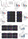
Figure 6.. Purine depletion by GCN2 inhibition triggers the Impaired Ribosome Biogenesis Checkpoint (IRBC), inducing p53 signaling.
(A) LNCaP cells were treated with GCN2iB (5 μM) or vehicle for 24 or 48 hours, mycophenolic acid (MPA; 1 μM) for 24 hours, or doxorubicin (Dox; 500 nM) for 24 hours. Lysates were prepared and analyzed by immunoblot using antibodies that recognize phosphorylated p53 (p-p53-Ser15), total p53, p21, MDM2, or actin. Molecular weight markers in kDa. The bar graphs to the right depict levels of the indicated protein normalized to the appropriate control (N = 3 biological replicates). Statistical significance was determined using one-way ANOVA with Tukey’s multiple comparisons. Error bars indicate SD. (B) Levels of purine nucleotides in LNCaP cells treated with vehicle, GCN2iB (5 μM), or MPA (1 μM) for 24 hours. Measurements are presented as picomoles (pmol) of nucleotide normalized to μg of total protein. Adenine nucleotide pools = ATP + ADP + AMP, and guanine nucleotide pools = GTP + GDP. Individual nucleotide measurements are shown in fig. S12C. Statistical significance was determined using a one-way ANOVA with Tukey’s multiple comparisons (N = 4 biological replicates). Error bars indicate SD. (C) Sucrose gradient analyses (-MgCl2) to measure levels of ribosomal subunits in LNCaP cells treated with vehicle, GCN2iB (5 μM), or MPA (1 μM) for 48 hours. Gradients were fractionated and monitored for absorbance at 254 nm. (D) Measurements of 3H-Uridine incorporation into 40S and 60S ribosomal subunits from panel (C). Statistical significance was determined using a one-way ANOVA with Tukey’s multiple comparisons (N = 3–5 biological replicates for (C and D)). Error bars indicate SD. (E) 47S pre-rRNA was measured by qRT-PCR in LNCaP cells treated with vehicle, GCN2iB (5 μM), or MPA (1 μM) for 48 hours. Statistical significance was determined using a one-way ANOVA with Tukey’s multiple comparisons (N = 4 biological replicates). Error bars indicate SD. (F and G) Immunofluorescent analysis of the nucleolar incorporation of 5-EU in (F) LNCaP cells treated with vehicle or GCN2iB (5 μM) for 48 hours, and (G) WT or GCN2 KO 22Rv1 cells, each pulsed with 5-EU for 1 hour, labelled with Cy-5 using Click-iT, and co-stained with Hoechst 33342 and the nucleolar marker fibrillarin. Statistical significance was determined using an unpaired two-tailed t-test (N = 8 to 10 optical fields from 3 biological replicates). Error bars indicate SD. (H) Representative images from (G) of 22Rv1 WT and GCN2 KO cells labeled with 5-EU and co-stained with fibrillarin and Hoechst 33342. (I) LNCaP cells were treated with vehicle or GCN2iB (5 μM) with or without EAAs or adenine (50 μM) for 24 or 48 hours, and cells were pulsed with 5-EU for 1 hour. The incorporation of 5-EU that was labeled with Cy-5 was monitored using Click-iT, and the cells were co-stained with SYTOX Green nucleic acid stain. The 5-EU and SYTOX Green signals were measured using a LI-COR instrument. Bar graphs indicate 5-EU intensity normalized to SYTOX Green, and statistical significance was determined using a two-way ANOVA with Tukey’s multiple comparisons (N = 5 to 6 biological replicates). Error bars indicate SD. (J to L) LNCaP cells were treated with vehicle for 48 hours, GCN2iB (5 μM) for 48 hours, or MPA (1 μM) for 24 hours. Whole-cell extracts (WCE) and input lysates (lysates that were cleared of ribosomes by ultracentrifugation) were analyzed by immunoblotting (J). Input lysates were immunoprecipitated with antibody to either MDM2 (K) or to RPL11 (L) and analyzed by immunoblotting. Molecular weight markers are in kDa. Blots are representative of two independent experiments. For all panels, *p ≤ 0.05, **p ≤ 0.001, ***p ≤ 0.001, and ****p ≤ 0.0001 by the test indicated.
Figure 7.
Figure 7.. Loss of GCN2 or p53 in PCa cells decreases ribosome biogenesis.
(A and B) LNCaP cells transfected with p53 siRNA or scrambled siRNA for 24 hours, followed by treatment with GCN2iB (5 μM), or vehicle for an additional 48 hours. Cell lysates were prepared and subjected to sucrose gradient centrifugation (-MgCl2), and fractions were monitored for absorbance at 254 nm. The 40S and 60S ribosomal subunits are indicated in the polysome profile. Measurements of 3H-Uridine incorporation into 40S and 60S ribosomal subunits are in (B). Statistical significance was determined using a one-way ANOVA with Tukey’s multiple comparisons (N = 3 biological replicates). Error bars indicate SD. (C) 47S pre-rRNA was measured by qRT-PCR in LNCaP WT or LNCaP p53 KO cells treated with vehicle or GCN2iB (5 μM) for 48 hours. Statistical significance was determined using a one-way ANOVA with Tukey’s multiple comparisons (N = 3–4 biological replicates). Error bars indicate SD. (D and E) Immunofluorescence analysis of nucleolar incorporation of 5-EU in WT or p53 KO LNCaP cells treated with vehicle or GCN2iB (5 μM) for 48 hours (top), and in WT or p53 KO 22Rv1 cells treated with vehicle or GCN2iB for 72 hours (bottom), each pulsed with 5-EU for 1 hour. 5-EU signal was quantified by modification with Cy-5 using Click-iT. Cells were co-stained with Hoechst 33342 and the nucleolar marker fibrillarin. Representative images of 22Rv1 WT and p53 KO cells treated with or without GCN2iB, labeled with 5-EU, and co-stained with fibrillarin and Hoechst 33342 are in (E). Statistical significance was determined using one-way ANOVA with Tukey’s multiple comparisons (N = 8–10 optical fields from 3 biological replicates). Error bars indicate SD. (F to H) DKO (Pten−/− Rb1−/−-) and TKO (Pten−/− Rb1−/− TP53−/−) organoids (N = 8 to 10 organoids) were treated with vehicle or GCN2iB (5 μM) for 5 days and pulsed with 5-EU for 1 hour (F). In panels (G) and (H), respectively, DKO and TKO organoids (N ≥ 12 organoids each) were also separately treated with vehicle or GCN2iB (5 μM) with or without EAAs or adenine (50 μM) for 5 days and pulsed with 5-EU for 1 hour. For panels (E to G), the 5-EU was labeled with Cy-5 using Click-iT, and organoids were stained with Hoechst 33342. The nucleolar 5-EU signal normalized to the organoid area (Hoechst 3342 signal) was quantified by immunofluorescence and is depicted in the bar graphs. Statistical significance was determined using a two-way ANOVA with Tukey’s multiple comparisons. Error bars indicate SD. For all panels, *p ≤ 0.05, **p ≤ 0.001, ***p ≤ 0.001, and ****p ≤ 0.0001 by the test indicated.
Figure 8.
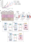
Figure 8.. Loss of p53 enhances sensitivity to GCN2 inhibition in the 22Rv1 xenograft model.
(A) Tumor volumes on the indicated days in male NSG mice bearing subcutaneous 22Rv1 WT or 22Rv1 p53 KO xenografts and treated with vehicle or 30 mg/kg GCN2iB twice daily for 5 days per week. N = 6 mice per group. Statistical significance was determined using a two-way repeated measures ANOVA with Sidak’s multiple comparisons. Error bars indicate the SEM. (B) Staining for cleaved caspase 3, senescence-associated β-galactosidase (SA-β-gal), or Ki-67 in the tumors from mice described in (A). Images are representative of 5 to 6 tumor sections, each from an independent mouse. Quantification for each stain is displayed as percent positive cells in the bar graphs. Statistical significance was determined using a one-way ANOVA with Tukey’s multiple comparisons. Error bars indicate SD. For all panels, * p ≤ 0.05, ** p ≤ 0.01, and *** p ≤ 0.001 by the test indicated. (C) Model depicting the response of p53 to GCN2 inhibition in PCa cells. Inhibition of GCN2 and the ISR disrupts amino acid homeostasis by reducing amino acid synthesis and uptake. A decrease in amino acid levels reduces purine pools, ultimately impairing ribosome biogenesis that triggers the Impaired Ribosome Biogenesis Checkpoint (IRBC). The IRBC stabilizes p53 levels and increases p53 signaling, which promotes G1 cell cycle arrest and senescence.
Figure 9.
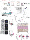
Figure 9.. In vivo CRISPRi screen reveals that GART inhibition sensitizes PCa cells to GCN2 inhibition.
(A) Schematic of 22Rv1 WT or 22Rv1 GCN2 KO CRISPRi screen in an in vivo tumor model. (B) Gene-level depletions are featured for 22Rv1 GCN2 KO tumors vs 22Rv1 WT tumors (data file S6). The average fold change for each gene is shown on the x-axis, and significance [adjusted p-value (−log10)] is presented on the y-axis. Significantly depleted genes (fold-change ≤ −2, p ≤ 0.05) in 22Rv1 GCN2 KO tumors are indicated in blue, and GART is highlighted in red. Information on all genes from the CRISPRi screen is shown in data file S6. (C) GSEA using the Reactome dataset of significantly depleted genes identified in the CRISPRi screen (data file S6). The plot indicates significantly enriched gene sets (p-value ≤ 0.05), and the bubble size indicates the number of significantly depleted genes corresponding to each gene set. (D) TM00298 organoids were treated with vehicle, GCN2iB (5 μM), pelitrexol (50 nM), or the combination of GCN2iB and pelitrexol for 10 days. Organoids were stained with Hoechst 33342 and Cytotox Red, and representative brightfield and immunofluorescence microscopy images are shown for each treatment regimen. Bar graphs show average area (mm2) and cell death as determined by Cytotox Red staining. Statistical significance was determined by one-way ANOVA with Tukey’s multiple comparisons (N ≥ 10 organoids). (E) Tumor volumes on the indicated days in male NSG mice (N = 6 to 8 mice per group) bearing subcutaneous xenografts of 22Rv1 WT or GCN2 KO 22Rv1 cells and treated with vehicle or 20 mg/kg pelitrexol every three days for a total of 21 days. Statistical significance was determined using a two-way repeated measures ANOVA with Sidak’s multiple comparisons. Error bars indicate SEM. (F) Staining for cleaved caspase 3, SA-β-gal, and Ki-67 are shown for tumors from (E). Quantification for each stain is displayed as percent positive cells in the bar graphs. Images are representative of 6 tumor sections, each from an independent mouse. Statistical significance was determined using a one-way ANOVA with Tukey’s multiple comparisons. Error bars indicate SD. For all panels, *p ≤ 0.05, **p ≤ 0.001, ***p ≤ 0.001, and ****p ≤ 0.0001 by the test indicated.

References

    1. Baird TD and Wek RC, Eukaryotic initiation factor 2 phosphorylation and translational control in metabolism. Adv Nutr, 2012. 3(3): p. 307–21. - PMC - PubMed
    1. Staschke KA and Wek RC, Adapting to cell stress from inside and out. Nat Cell Biol, 2019. 21(7): p. 799–800. - PubMed
    1. Wek RC, Anthony TG, and Staschke KA, Surviving and Adapting to Stress: Translational Control and the Integrated Stress Response. Antioxid Redox Signal, 2023. 39(4–6): p. 351–373. - PMC - PubMed
    1. Wek RC and Staschke KA, How do tumours adapt to nutrient stress? EMBO J, 2010. 29(12): p. 1946–7. - PMC - PubMed
    1. Siegel RL, et al., Cancer statistics, 2023. CA Cancer J Clin, 2023. 73(1): p. 17–48. - PubMed

Publication types