. 2024 Nov 26;31(10-11):a054026.
doi: 10.1101/lm.054026.124. Print 2024 Oct-Nov.

Lateral entorhinal cortex neurons that project to nucleus accumbens mediate contextual associative memory

Affiliations

Lateral entorhinal cortex neurons that project to nucleus accumbens mediate contextual associative memory

Hayley M Kuhn et al. Learn Mem. .

Abstract

The lateral entorhinal cortex (LEC) contains glutamatergic projections that innervate the nucleus accumbens (NAc) and may be involved in the encoding of contextual associations with both positive and negative valences, such as those encountered in drug cues or fear conditioning. To determine whether LEC-NAc neurons are activated by the encoding and recall of contexts associated with cocaine or footshock, we measured c-fos expression in these neurons and found that LEC-NAc neurons are activated in both contexts. Specifically, activation patterns of the LEC-NAc were observed in a novel context and reexposure to the same context, highlighting the specific role for LEC-NAc neurons in encoding rather than the valence of a specific event-related memory. Using a combination of circuit-specific chemogenetic tools and behavioral assays, we selectively inactivated LEC-NAc neurons in mice during the encoding and retrieval of memories of contexts associated with cocaine or footshock. Chemogenetic inactivation of LEC-NAc neurons impaired the formation of both positive and negative context-associated memories without affecting the retrieval of an established memory. This finding suggests a critical role for this circuit in the initial encoding of contextual associations. In summary, LEC-NAc neurons facilitate the encoding of contextual information, guiding motivational behaviors without directly mediating the hedonic or aversive properties of these associations.


Figures

Figure 1.
Figure 1.
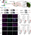
LEC-NAc neurons are activated by novel conditioning contexts. (A) Circuit-tagging method to label LEC-NAc neurons with GFP. Cre-dependent Rosa26eGFP-L10a male and female mice received intracranial infusion of a retrograde Cre virus (HSVrg-hEf1α-Cre) to drive expression of GFP in LEC-NAc neurons. (B) Experimental approach schematic and timeline of brain extraction for c-fos immunohistochemistry. (C) Representative 20 fluorescent images from LEC showing GFP + cells (left, magenta), c-fos + cells (middle, cyan), and merged image (right). White arrows indicate overlap and denote c-fos + GFP + LEC-NAc neurons. (D) Quantification of cell counts in each ROI (per mm2) expressing GFP (left), c-fos (middle), and colabeled (right) in Home (n = 3 mice), Sal CPP (n = 5 mice), and Coc CPP groups (n = 5 mice). (E) Quantification of cell counts in each ROI (per mm2) expressing GFP (left), c-fos (middle), and colabeled (right) in Home (n = 3 mice; same control used in D), Context (n = 4 mice), and Context + FS groups (n = 5 mice). One-way ANOVA followed by Holm–Sidak post hoc comparisons; (*) P < 0.05, (**) P < 0.01, (***) P < 0.001.
Figure 2.
Figure 2.
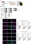
LEC-NAc neurons are activated by retrieval of context. (A) Circuit-tagging method to label LEC-NAc neurons with GFP. Cre-dependent Rosa26eGFP-L10a male and female mice received intracranial infusion of a retrograde Cre virus (HSVrg-hEf1α-Cre) to drive expression of GFP in LEC-NAc neurons. (B) Experimental approach schematic and timeline of brain extraction for c-fos immunohistochemistry. (C) Representative 20 fluorescent images from LEC showing GFP + cells (left, magenta), c-fos + cells (middle, cyan), and merged image (right). White arrows indicate overlap and denote c-fos + GFP + LEC-NAc neurons. (D) Quantification of cell counts in each ROI (per mm2) expressing GFP (left), c-fos (middle), and colabeled (right) in Home (n = 6 mice), No CPP Test (n = 5 mice), Sal CPP Test (n = 5 mice), and Coc CPP Test groups (n = 5 mice). (E) Quantification of cell counts in each ROI (per mm2) expressing GFP (left), c-fos (middle), and colabeled (right) in Home (n = 6 mice; same control used in D), No CFC Test (n = 6 mice), Context Test (n = 5 mice), and CFC Test groups (n = 6 mice). One-way ANOVA followed by Holm–Sidak post hoc comparisons; (*) P < 0.05, (**) P < 0.01, (***) P < 0.001, (****) P < 0.0001.
Figure 3.
Figure 3.
LEC-NAc neurons are necessary for the encoding, but not the retrieval of a cocaine-context association. (A) Circuit-specific DREADD approach to inhibit LEC-NAc neurons during cocaine-context conditioning. (B) Pretest time spent within the prospective cocaine-paired chamber (Coc), prospective saline-paired chamber (Sal), and center chamber in mCherry-expressing controls (mCh) and Gi-DREADD-expressing mice (Gi). There were no differences between Coc and Sal chambers in each group. Two-way RM ANOVA, P > 0.05. (C) Pretest preference (Coc time minus Sal time) within each group. There were no pretest differences between groups. t-test, P > 0.05. (D) Posttest chamber time in each group. mCh mice spent more time in Coc compared to Sal; however, Gi mice spent an equivalent amount of time in both chambers. Two-way RM ANOVA followed by Holm–Sidak post hoc comparisons, (****) P < 0.0001. (E) Posttest preference within each group. Gi mice had impaired preference for the Coc chamber (relative to Sal) compared to mCh controls. t-test, P < 0.05. (F) Circuit-specific DREADD approach to inhibit LEC-NAc neurons during cocaine-context posttest. (G) Pretest time spent within the prospective Coc, Sal, and center chambers in mCh controls (mCh-CNO), Gi mice that receive vehicle during posttest (Gi-DMSO), and Gi mice that receive CNO during posttest (Gi-CNO). There were no differences between Coc and Sal chambers in each group. Two-way RM ANOVA, P > 0.05. (H) Pretest preference within each group. There were no pretest differences between groups. t-test, P > 0.05. (I) Posttest chamber time in each group. All groups spent more time in Coc compared to Sal. Two-way RM ANOVA, (*) P < 0.05. (J) Posttest preference within each group. There were no differences between groups. One-way ANOVA, P > 0.05.
Figure 4.
Figure 4.
LEC-NAc neurons are necessary for the encoding, but not the retrieval of contextual fear memory. (A) Circuit-specific DREADD approach to inhibit LEC-NAc neurons during footshock-context conditioning. (B) Contextual fear memory test freezing time across 1 min bins in mCherry controls (mCh) and Gi-DREADD mice (Gi). Gi mice had decreased freezing across the test bins compared to mCh controls. Two-way RM ANOVA followed by Holm–Sidak post hoc comparisons, (*) P < 0.05. (C) Percent of freezing time for the entire test within each group. Gi mice spent less % time freezing compared to mCh mice. t-test, (*) P < 0.05. (D) Circuit-specific DREADD approach to inhibit LEC-NAc neurons during contextual fear memory test. (E) Contextual fear memory test freezing time across 1 min bins in mCh controls and Gi mice. There were no differences between groups. Two-way RM ANOVA, P > 0.05. (F) Percent of freezing time for the entire test within each group. There were no differences between groups. t-test, P > 0.05.

References

    1. Allichon M-C, Ortiz V, Pousinha P, Andrianarivelo A, Petitbon A, Heck N, Trifilieff P, Barik J, Vanhoutte P. 2021. Cell-type-specific adaptions in striatal medium-sized spiny neurons and their roles in behavioral responses to drugs of abuse. Front Synaptic Neurosci 13: 799274. 10.3389/fnsyn.2021.799274 - DOI - PMC - PubMed
    1. Aylward RL, Totterdell S. 1993. Neurons in the ventral subiculum, amygdala and entorhinal cortex which project to the nucleus accumbens: their input from somatostatin-immunoreactive boutons. J Chem Neuroanat 6: 31–42. 10.1016/0891-0618(93)90005-O - DOI - PubMed
    1. Baldi E, Bucherelli C. 2014. Entorhinal cortex contribution to contextual fear conditioning extinction and reconsolidation in rats. Neurobiol Learn Memory 110: 64–71. 10.1016/j.nlm.2014.02.004 - DOI - PubMed
    1. Baldi E, Liuzzo A, Bucherelli C. 2013. Fimbria–fornix and entorhinal cortex differential contribution to contextual and cued fear conditioning consolidation in rats. Physiol Behav 114115: 42–48. 10.1016/j.physbeh.2013.03.011 - DOI - PubMed
    1. Bariselli S, Fobbs WC, Creed MC, Kravitz AV. 2019. A competitive model for striatal action selection. Brain Res 1713: 70–79. 10.1016/j.brainres.2018.10.009 - DOI - PMC - PubMed

MeSH terms

LinkOut - more resources