. 2024 Nov 26;19(1):122.
doi: 10.1186/s13062-024-00566-y.

YTHDF2 promotes anaplastic thyroid cancer progression by activating the DDIT4/AKT/mTOR signaling pathway

Affiliations

YTHDF2 promotes anaplastic thyroid cancer progression by activating the DDIT4/AKT/mTOR signaling pathway

Bao Dai et al. Biol Direct. .

Abstract

Background: RNA methylation, an important reversible post-transcriptional modification in eukaryotes, has emerged as a prevalent epigenetic alteration. However, the role of the m6A reader YTH domain family 2 (YTHDF2) has not been reported in anaplastic thyroid cancer (ATC) and its biological mechanism is unclear.

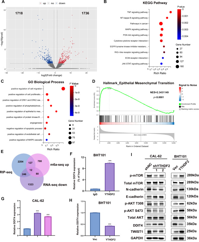
Methods: The relationship between YTHDF2 expression and ATC was determined using data sets and tissue samples. A range of analytical techniques were employed to investigate the regulatory mechanism of YTHDF2 in ATC, including bioinformatics analysis, m6A dot-blot analysis, methylated RNA immunoprecipitation sequencing (MeRIP-seq), RNA immunoprecipitation (RIP) assays, RNA sequencing, RNA stability assays and dual luciferase reporter gene assays. In vitro and in vivo assays were also conducted to determine the contribution of YTHDF2 to ATC development.

Results: YTHDF2 expression was significantly increased in ATC. The comprehensive in vitro and in vivo experiments demonstrated that YTHDF2 knockdown significantly attenuated ATC proliferation, invasion, migration, and apoptosis promotion, whereas YTHDF2 overexpression yielded the opposite trend. Mechanistically, RNA-seq, MeRIP-seq and RIP-seq analysis, and molecular biology experiments demonstrated that YTHDF2 accelerated the degradation of DNA damage-inducible transcript 4 or regulated in DNA damage and development 1 (DDIT4, or REDD1) mRNA in an m6A-dependent manner, which in turn activated the AKT/mTOR signaling pathway and induced activation of epithelial-mesenchymal transition (EMT), thereby promoting ATC tumor progression.

Conclusions: This study is the first to demonstrate that elevated YTHDF2 expression levels suppress DDIT4 expression in an m6A-dependent manner and activate the AKT/mTOR signaling pathway, thereby promoting ATC progression. YTHDF2 plays a pivotal role in ATC progression, and it may serve as a promising therapeutic target in the future.

Keywords: AKT; Anaplastic thyroid cancer; YTHDF2; m6A; mTOR.


Conflict of interest statement

Declarations. Ethics approval and consent to participate: The study was approved by the Ethical Review Committee of the Guangdong Provincial People’s Hospital and was conducted in accordance with the Declaration of Helsinki (Project No. KY2023-306). All animals were housed and handled according to the guidelines of the Ethical Review Committee of Guangdong Provincial People’s Hospital (acceptance No. S2023-357-02.20160006). Consent for publication: Not applicable. Competing interests: The authors declare no competing interests.

Figures

Fig. 1
Fig. 1
YTHDF2 and m6A expression levels in ATC. A The YTHDF2 expression pattern were analyzed in 512 thyroid tumors and 338 normal controls (UCSC XENA database). The Mann-Whitney test was used for statistical analysis. B Representative IHC staining images of YTHDF2 expression in ATC, FTC and normal thyroid tissues. C YTHDF2 protein expression levels in normal thyroid, FTC and ATC cell lines. D Immunofluorescence detection of the subcellular localization of YTHDF2, scale bar = 10 μm. E, F YTHDF2 was knocked down and overexpressed in CAL-62 and BHT101 cells, respectively, and the efficiency was verified by qPCR and WB. GAPDH was the internal reference. G m6A RNA dot-blot assay. m6A level changes were detected in CAL-62 and BHT101 cell lines at different total RNA concentrations (200 ng, 400 ng). Methylene blue staining was used as loading control. H Immunofluorescence detection of m6A levels in CAL-62 and BHT101 cell lines. Scale bar = 20 μm. Error bars represent the SD obtained from at least three independent experiments. *P < 0.05, **P < 0.01, ***P < 0.001
Fig. 2
Fig. 2
YTHDF2 promoted ATC malignant phenotype progression. A-C Effect of YTHDF2 on ATC proliferation detected by clonal colony formation assay, EdU assay, and CCK-8 assay. D F-actin fluorescent staining measurement of the effect of YTHDF2 overexpression and knockdown on cell cytoskeleton. Representative images of the F-actin fluorescent staining are shown. Scale bar = 10 μm. E, F Trans-well assay and wound healing assay to verify the effect of YTHDF2 knockdown and overexpression on cell migration ability. G Flow cytometry assay to confirm the apoptosis analysis induced by YTHDF2 knock-down and overexpression. Error bars represent the SD from at least three independent experiments. *P < 0.05, **P < 0.01, ***P < 0.001
Fig. 3
Fig. 3
YTHDF2 facilitated the ATC progression by activating the AKT-mTOR pathway and EMT by suppressing DDIT4 expression. A Fold change in differential expression of genes in different subgroups of MeRIP (shNC, shYTHDF2) was represented by log2(foldchange) as the horizontal coordinate. Red color represents up-regulated significant differential expression (1736 genes) and blue color represents down-regulated significant differential expression (1718 genes). B, C KEGG analysis and GO-BP enrichment analysis were performed on the differential genes (|log2FC| > 0.5, P < 0.05). D GSEA revealing that the YTHDF2-related differential genes are significantly enriched in the EMT pathway. E Venn diagram depicting the intersecting genes from the MeRIP-seq, RIP-seq and mRNA-seq correlated genes. Twelve common genes were screened out. F RIP-qPCR confirmation of the DDIT4 mRNA enrichment by YTHDF2 in BHT cell line. G-I qPCR and WB verification of DDIT4 expression levels. I Validation of the effect of YTHDF2 on AKT/mTOR and EMT pathway by WB. Error bars represent the SD from at least three independent experiments; *P < 0.05, **P < 0.01, and ***P < 0.001
Fig. 4
Fig. 4
YTHDF2 mediated the DDIT4 mRNA degradation in an m6A-dependent manner. A IGV plots showed the m6A and YTHDF2 binding site and the change of RNA-seq peaks at DDIT4 mRNAs in CAL-62. B The sequence represents the fragments captured in MeRIP-seq, which was co-localized with predicted sites. C The m6A peaks motifs are RRACH (R = G/A, H = A/C/U). D and E Relative luciferase activity of DDIT4 5’UTR with wild-type or mutated m6A sites after YTHDF2 knockdown and overexpression in CAL-62 and BHT101 cells. Renilla luciferase activity was measured and normalized to firefly luciferase activity. F The total RNA m6A level after DAA treatment was detected by m6A RNA dot blot assay and compared with DMSO treatment, with methylene blue staining as a loading control. G qPCR was used for the detection of DDIT4 mRNA levels after DAA treatment. H MeRIP-RT-qPCR detection of m6A level alterations of DDIT4 after METTL3 knockdown in CAL-62 cells. I RT-qPCR analysis of the decay rate of DDIT4 mRNA after actinomycin D (5 µg/mL) treatment in CAL-62 and BHT101 cells with YTHDF2knockdown or overexpression. Error bars represent the SD from at least three independent experiments. *P < 0.05, **P < 0.01, ***P < 0.001
Fig. 5
Fig. 5
YTHDF2 promoted tumor growth and metastasis in vivo. A, B The tumor growth curve of xenografts was plotted by measuring the tumor size (width^2 × length × 0.52) with caliper. The tumor size at the endpoint in the above group was analyzed by Student’s t-test. The subcutaneous tumor models were observed after 28 days, the anatomized subcutaneous tumor xenografts were weighed and analyzed with Student’s t-test. C Representative IHC staining micrographs of YTHDF2 in tumor xenografts were conducted. D. Proteins were extracted from animal subcutaneous tumors for WB detection of EMT-associated proteins. E. The BALB/c nude mice injected with cells via tail vein were imaged at week 4 to evaluate the entire metastasis by the IVIS. F. H&E staining of multiple metastatic organs was performed to identify the metastatic loci. Error bars represent the SD obtained from at least three independent experiments.*P < 0.05, **P < 0.01, ***P < 0.001
Fig. 6
Fig. 6
The tumor suppressor role of DDIT4 in ATC. A To verify the transfection efficiency of DDIT4 and detect the expression levels of AKT/mTOR and EMT-related proteins by WB. GAPDH was the internal reference. B, C EdU and CCK-8 assay evaluation of the proliferative capacity of cells after DDIT4 overexpression. D, E Trans-well and wound healing assay evaluation of cell migration upon DDIT4 overexpression. F Flow cytometry assay evaluation of the apoptosis induced by DDIT4 overexpression. G Schematic diagram representing the results of this study. Error bars represent the SD from at least three independent experiments. *P < 0.05, **P < 0.01, ***P < 0.001

Similar articles

References

    1. Bray F, Laversanne M, Sung H, Ferlay J, Siegel RL, Soerjomataram I, Jemal A. Global cancer statistics 2022: GLOBOCAN estimates of incidence and mortality worldwide for 36 cancers in 185 countries. CA Cancer J Clin 2024, 74(3):229-263. - PubMed
    1. Han B, Zheng R, Zeng H, Wang S, Sun K, Chen R, Li L, Wei W, He J. Cancer incidence and mortality in China, 2022. J Natl Cancer Cent. 2024;4(1):47–53. - PMC - PubMed
    1. Molinaro E, Romei C, Biagini A, Sabini E, Agate L, Mazzeo S, Materazzi G, Sellari-Franceschini S, Ribechini A, Torregrossa L, et al. Anaplastic thyroid carcinoma: from clinicopathology to genetics and advanced therapies. Nat Rev Endocrinol. 2017;13(11):644–60. - PubMed
    1. Boucai L, Zafereo M, Cabanillas ME. Thyroid Cancer: a review. JAMA. 2024;331(5):425–35. - PubMed
    1. Perrier ND, Brierley JD, Tuttle RM. Differentiated and anaplastic thyroid carcinoma: major changes in the American Joint Committee on Cancer eighth edition cancer staging manual. CA Cancer J Clin. 2018;68(1):55–63. - PMC - PubMed

MeSH terms

LinkOut - more resources