. 2024 Nov 13;11(11):1142.
doi: 10.3390/bioengineering11111142.

Upper Crossed Syndrome and Scapulae Upper-Trapping: A Mesotherapy Protocol in Cervicoscapulobrachial Pain-The 8:1 Block

Affiliations

Upper Crossed Syndrome and Scapulae Upper-Trapping: A Mesotherapy Protocol in Cervicoscapulobrachial Pain-The 8:1 Block

Luyddy Pires et al. Bioengineering (Basel). .

Abstract

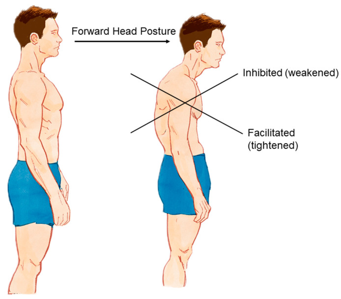
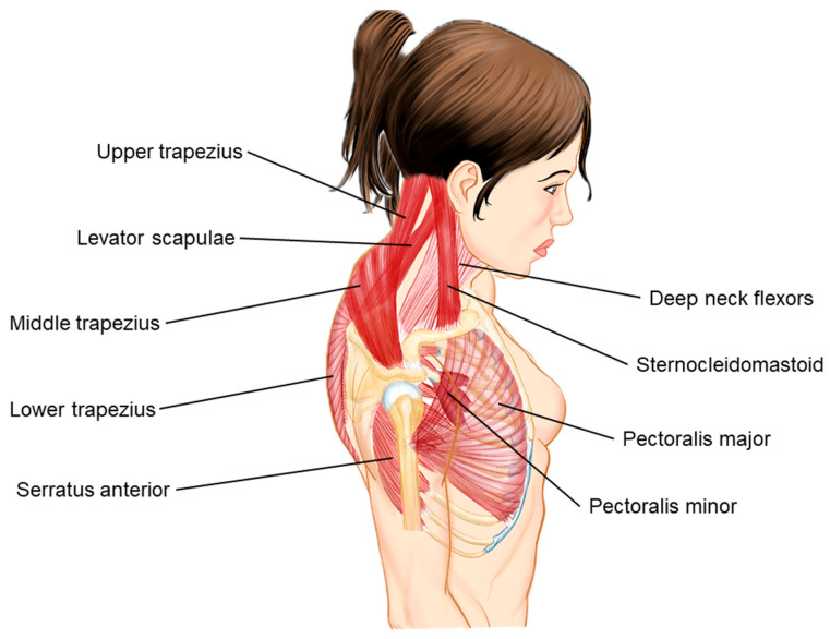
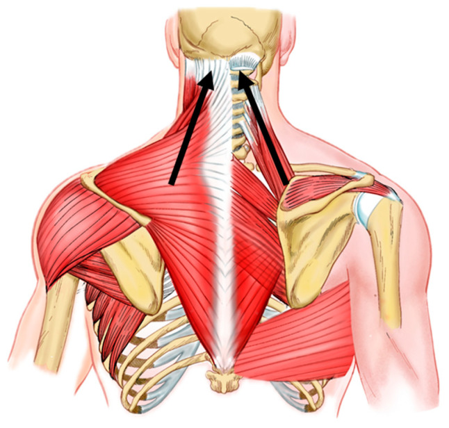
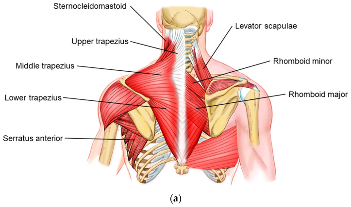
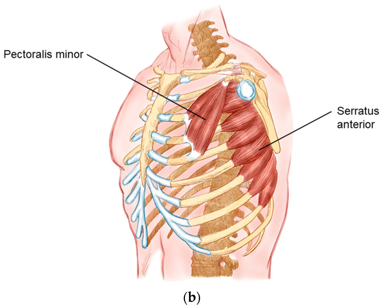
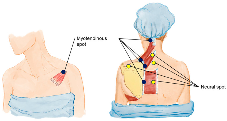
Upper Crossed Syndrome (UCS), described by Vladimir Janda, is characterized by postural changes involving the cervical spine and trunk, leading to biomechanical limitations and cervicoscapulobrachial pain. This study proposes a mesotherapy protocol, termed the 8:1 block, to address cervicoscapulobrachialgia by targeting the scapulae and associated musculature. The scapula, central to shoulder girdle kinematics, often exhibits dyskinesis and muscular imbalances, notably the pattern referred to as scapular upper trapping (SUT). SUT involves scapular elevation, medial rotation, and shoulder protraction, contributing to cervicobrachial pain. The protocol includes a comprehensive assessment of muscle tone changes and biomechanical considerations, highlighting the importance of the scapula in upper limb movement and posture. Key anatomical changes involve tightened upper trapezius, levator scapulae, and pectoralis minor muscles, with weakened middle trapezius and serratus anterior. The mesotherapy approach targets these imbalances through specific injection points to alleviate muscle tension and correct postural deviations. Case studies from our clinic demonstrate the protocol's effectiveness in reducing pain and restoring scapular biomechanics. Patients reported significant improvements in pain relief and functional outcomes, underscoring the clinical utility of the 8:1 block in treating cervicoscapulobrachialgia. This protocol offers a feasible, cost-effective intervention that enhances the efficacy of traditional therapeutic exercises by addressing underlying muscular and biomechanical dysfunctions. In conclusion, the 8:1 block mesotherapy protocol provides a novel approach to managing cervicoscapulobrachial pain by focusing on scapular biomechanics and muscle tension. Further studies are needed to validate these findings and refine the protocol for broader clinical application.

Keywords: cervicoscapulobrachialgia; mesotherapy protocol; scapular dyskinesis; scapular upper trapping; upper crossed syndrome.


Conflict of interest statement

The authors declare no conflicts of interest.

Figures

Figure 1
Figure 1
Postural change of the UCS leading to FHP. A normal posture (left) compared with the altered posture associated with UCS (right), characterized by FHP and a cross pattern of muscle imbalances. The inhibited (weakened) muscles are indicated by the upper arrows, and the facilitated (tightened) muscles are shown by the lower arrows. These postural changes contribute significantly to cervicoscapulobrachial pain and dysfunction.
Figure 2
Figure 2
Muscle imbalances in UCS. The labeled muscles, related to the UCS, include the upper trapezius, levator scapulae, middle trapezius, lower trapezius, serratus anterior, deep neck flexors, sternocleidomastoid, pectoralis major, and pectoralis minor. The image highlights the typical postural distortion seen in UCS, where the upper trapezius and levator scapulae (dark red) are often overactive and tight, while the deep neck flexors and lower trapezius are typically weak (light red). These imbalances build the dysfunction and pain in the cervicoscapulobrachial region.
Figure 3
Figure 3
Tension lines in UCS. The usual vectors of tension in UCS are indicated by black arrows. Upper trapezius and levator scapulae muscles are often tightened and overactive in UCS. These tension lines represent the typical patterns of muscle imbalance and postural distortion seen in UCS, contributing to cervicoscapulobrachial pain and leading to the SUT definition. Understanding these tension lines is crucial for effectively targeting therapeutic interventions.
Figure 4
Figure 4
(a) Muscles involved in UCS and SUT. This illustration depicts the key muscles involved in the pathology of UCS and scapulae upper-trapping. The labeled muscles include the sternocleidomastoid, levator scapulae, upper trapezius, middle trapezius, lower trapezius, serratus anterior, supraspinatus, rhomboid minor, rhomboid major, and latissimus dorsi. The image highlights both the superficial and deep muscles contributing to the characteristic muscle imbalances and postural distortions seen in UCS, which are often associated with cervicoscapulobrachial pain. This context is crucial for understanding the mesotherapy protocol proposed in the 8:1 block treatment approach. (b) Muscles Involved in Scapular Movement and Stabilization. This illustration highlights the pectoralis minor and serratus anterior muscles, which play crucial roles in scapular movement and stabilization. The tightening of the pectoralis minor is often implicated in the protraction and internal rotation of the scapula, contributing to the muscle imbalances seen in UCS. The serratus anterior is essential for proper scapular motion and stability, and its weakness is commonly observed in UCS.
Figure 4
Figure 4
(a) Muscles involved in UCS and SUT. This illustration depicts the key muscles involved in the pathology of UCS and scapulae upper-trapping. The labeled muscles include the sternocleidomastoid, levator scapulae, upper trapezius, middle trapezius, lower trapezius, serratus anterior, supraspinatus, rhomboid minor, rhomboid major, and latissimus dorsi. The image highlights both the superficial and deep muscles contributing to the characteristic muscle imbalances and postural distortions seen in UCS, which are often associated with cervicoscapulobrachial pain. This context is crucial for understanding the mesotherapy protocol proposed in the 8:1 block treatment approach. (b) Muscles Involved in Scapular Movement and Stabilization. This illustration highlights the pectoralis minor and serratus anterior muscles, which play crucial roles in scapular movement and stabilization. The tightening of the pectoralis minor is often implicated in the protraction and internal rotation of the scapula, contributing to the muscle imbalances seen in UCS. The serratus anterior is essential for proper scapular motion and stability, and its weakness is commonly observed in UCS.
Figure 5
Figure 5
Thermography applied to UCS diagnosis. Thermography shows hyper or hypo radiation spots. Inflammatory spots are often hypervascularized and the increased blood flow leads to hypercaptation in the images. These images correlate the hypercaptation spots on the scapula levator insertion, medium trapezius and rhomboid minor.
Figure 6
Figure 6
Simetograph and its application to UCS diagnosis. The simetograph is a simple device, a squared background that facilitates the visual identification of postural asymmetries. The images show a typical FHP and shoulder asymmetry (right shoulder higher).
Figure 7
Figure 7
Myotendinous and neural injection spots for UCS. This illustration identifies the specific myotendinous and neural injection spots targeted in the mesotherapy protocol for treating UCS. Yellow dots indicate neural spots, essential for releasing fasciae entrapments, modulating neural inputs and reducing pain. Blue dots highlight additional target areas on the myotendinous junctions of muscles affected by UCS. These targeted injection spots are integral to the 8:1 block treatment approach, aiming to correct muscle imbalances and alleviate CSB pain associated with UCS.
Figure 8
Figure 8
Nerve anatomy relevant to UCS. Schematic representation of the key nerves involved in the pathology of UCS. The labeled nerves include the suprascapular nerve, the accessory nerve (superficial), and the dorsal scapular nerve (deep). These nerves innervate critical muscles that are often affected in UCS, contributing to muscle imbalances, weakness, and pain in the cervicoscapulobrachial region.
Figure 9
Figure 9
(a) Mesotherapy injection protocol for the cervicoscapulobrachial region in UCS, posterior view. (b) Anterior view.

Similar articles

References

    1. Janda V. Janda Compendium. Volume II. O.P.T.P.; Plymouth, MN, USA: 1997. pp. 7–13.
    1. Stevens G.T. The Pose of the Body as Related to the Type of the Cranium and the Direction of the Visual Plane. Buffalo Med. J. 1901;40:576–580. - PMC - PubMed
    1. Schwartz L. A resume, with comments, of the available literature relating to posture. Public Health Rep. 1927;42:1219–1224. doi: 10.2307/4578305. - DOI
    1. Sheikhhoseini R., Shahrbanian S., Sayyadi P., O’Sullivan K. Effectiveness of Therapeutic Exercise on Forward Head Posture: A Systematic Review and Meta-analysis. J. Manip. Physiol. Ther. 2018;41:530–539. doi: 10.1016/j.jmpt.2018.02.002. - DOI - PubMed
    1. Iannotti J.P., Parker R.D. The Netter Collection of Medical Illustrations (Frank H. Netter, MD). Musculoskeletal System, Part 1: Upper Limb. 2nd ed. Elsevier; Amsterdam, The Netherlands: 2013. pp. 2–3.

LinkOut - more resources