Review
. 2024 Oct 31;13(11):1330.
doi: 10.3390/antiox13111330.

Mitochondrial Reactive Oxygen Species Dysregulation in Heart Failure with Preserved Ejection Fraction: A Fraction of the Whole

Affiliations
Review

Mitochondrial Reactive Oxygen Species Dysregulation in Heart Failure with Preserved Ejection Fraction: A Fraction of the Whole

Caroline Silveira Martinez et al. Antioxidants (Basel). .

Abstract

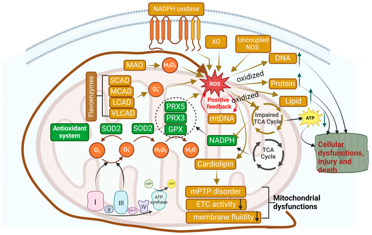
Heart failure with preserved ejection fraction (HFpEF) is a multifarious syndrome, accounting for over half of heart failure (HF) patients receiving clinical treatment. The prevalence of HFpEF is rapidly increasing in the coming decades as the global population ages. It is becoming clearer that HFpEF has a lot of different causes, which makes it challenging to find effective treatments. Currently, there are no proven treatments for people with deteriorating HF or HFpEF. Although the pathophysiologic foundations of HFpEF are complex, excessive reactive oxygen species (ROS) generation and increased oxidative stress caused by mitochondrial dysfunction seem to play a critical role in the pathogenesis of HFpEF. Emerging evidence from animal models and human myocardial tissues from failed hearts shows that mitochondrial aberrations cause a marked increase in mitochondrial ROS (mtROS) production and oxidative stress. Furthermore, studies have reported that common HF medications like beta blockers, angiotensin receptor blockers, angiotensin-converting enzyme inhibitors, and mineralocorticoid receptor antagonists indirectly reduce the production of mtROS. Despite the harmful effects of ROS on cardiac remodeling, maintaining mitochondrial homeostasis and cardiac functions requires small amounts of ROS. In this review, we will provide an overview and discussion of the recent findings on mtROS production, its threshold for imbalance, and the subsequent dysfunction that leads to related cardiac and systemic phenotypes in the context of HFpEF. We will also focus on newly discovered cellular and molecular mechanisms underlying ROS dysregulation, current therapeutic options, and future perspectives for treating HFpEF by targeting mtROS and the associated signal molecules.

Keywords: cardiac diastolic dysfunction; cardiovascular disease; heart failure; heart failure with preserved ejection fraction (HFpEF); mitochondrial; mitochondrial dysfunction; oxidative stress; reactive oxygen species; redox signal.


Conflict of interest statement

The authors declare no conflicts of interest.

Figures

Figure 1
Figure 1
Pathophysiology of HFpEF. Multiple cardiovascular (CV) and non-CV comorbidities sequentially cause chronic and low-grade systemic inflammation, mitochondrial dysfunction, and ROS generation. These pathological alterations consequently trigger pathological remodeling and functional changes in the heart, which in turn increase LV stiffness, eventually causing HFpEF. HFpEF, heart failure with preserved ejection fraction; LV, left ventricular; NO, nitric oxide; ROS, reactive oxygen species. Red upwards and downwards arrows indicate increase and decrease, respectively. Diagram was created with URL (BioRender.com) (accessed on 27 October 2024).
Figure 2
Figure 2
Mitochondrial metabolism in cardiac hemostasis. During cardiac hemostasis and under physiological conditions, the mitochondria in the heart produce 95% ATP by metabolizing a variety of fuels including fatty acids, glucose, lactate, ketones, pyruvate, and amino acids, primarily by mitochondrial oxidative phosphorylation (OXPHOS) through electron transport chain (ETC, or mitochondrial complex I–V). Tricarboxylic acid (TCA) cycle produces multiple metabolites (particularly NADH and FADH2), which enter ETC and function as electron carriers for ATP production. Additionally, 5% ATP is generated by anaerobic glycolysis. Diagram was created with URL (BioRender.com (accessed on 30 October 2024)).
Figure 3
Figure 3
Mitochondrial metabolism in HFrEF and HFpEF. A healthy heart uses fatty acids (FAs) as its primary energy fuel to produce ATP through fatty acid oxidation (FAO), while glucose becomes the main energy fuel for ATP generation in heart failure via glucose oxidation. Although impaired mitochondrial oxidative phosphorylation (OXPHOS) is a common feature for both HFrEF and HFpEF, evidence suggests that the failing heart in HFpEF attempts to compensate for the impaired OXPHOS by increasing BCAA (branched chain amino acid) rather than glucose oxidation. The latter is predominately observed in HFrEF. Multiple molecules (such as Sirt3, NAD+, ketone body β-hydroxybutyrate, SGLT2 inhibitor) have been reported as the key regulators for mitochondrial metabolism, which could be the novel therapeutics for HFpEF. Interestingly, PCSK9 (proprotein convertase subtilisin/kexin type 9) genetic deficiency may cause HFpEF by impairing FAO, raising caution for the clinical applications of PCSK9 inhibitors. Red upwards and downwards arrows indicate increase and decrease, respectively. Diagram was created with URL (BioRender.com) (accessed on 27 October 2024).
Figure 4
Figure 4
Mitochondrial quality control (MQC). MQC involved in mitochondrial biogenesis (new mitochondria generation), dynamics (fission and fusion), and mitophagy (degradation of the damaged or dysfunctional mitochondria) is a critical mechanism for protecting mitochondria and cellular functions. Decreased mitochondrial biogenesis, increased mitochondrial fission, and impaired mitophagy underscore HFpEF pathogenesis. Red upwards and downwards arrows indicate increase and decrease, respectively. Diagram was created with URL (BioRender.com) (accessed on 27 October 2024).
Figure 5
Figure 5
Mitochondrial ROS generation and potential detrimental effects. Although ROS can be generated by other cellular oxidative systems such as xanthine oxidase (XO), NADPH oxidase (Noxs), and uncoupled NOS, mitochondrial ROS (mtROS) generated from complexes I and III are the major source for cellular ROS. Cells including cardiomyocytes have an efficient antioxidant system situated within the mitochondrial matrix, neutralizing the “excessive” mtROS to fine-tuning cellular redox signaling. Imbalanced redox signaling generates excessive mtROS, which in turn oxidize DNAs, proteins, lipids, and mtDNAs and alter their corresponding functions, thereby causing a variety of cellular dysfunctions, injuries, and even death. Green upwards and downwards arrows indicate increase and decrease, respectively. Diagram was created with URL (BioRender.com) (accessed on 27 October 2024).
Figure 6
Figure 6
Mitochondrial ROS in mitohormesis and mitochondrial dysfunction in cardiomyocytes (CMs). Fine-tuning regulation of mitochondrial ROS (mtROS) is critical for cellular functions. While mitohormesis with low-to-mild levels of mtROS could activate protective pathways, improve cellular resilience against stress, and increase lifespan, excessive mtROS have detrimental effects on CMs such as hypertrophy, fibrosis, and apoptosis, eventually causing HFpEF. Green upwards and downwards arrows indicate increase and decrease, respectively. Diagram was created with URL (BioRender.com) (accessed on 27 October 2024).
Figure 7
Figure 7
Mitochondrial ROS in HFpEF. In HFpEF, there is a bidirectional crosstalk between mitochondria metabolic stress and inflammation, namely “mito-inflammation”. Excessive levels of mtROS initiate a vicious cycle of increased mtROS production, mitochondrial damage, and inflammation. Increased mtROS promotes the release of mtDNA and other mtDAMPs, which in turn activate proinflammatory signaling pathways, amplifying inflammation and further increasing mtROS generation. In HFpEF with diabetes, high levels of glucose and increased mtROS trigger glycoxidative stress, a detrimental positive feedback cycle of malignant AGE and ROS accumulation. The resulting mito-inflammation causes endothelial dysfunction, vasodilation impairment, and capillary rarefaction, all contributing to HFpEF pathogenesis. Moreover, the changed oxidative substrates, increased ROS, and defective mitochondria due to metabolic disruption and system inflammation also cause energy depletion, underscoring their role in endothelial dysfunction and HFpEF. Green upwards and downwards arrows indicate increase and decrease, respectively. Diagram was created with URL (BioRender.com) (accessed on 27 October 2024).

Similar articles

Cited by

References

    1. McDonagh T.A., Metra M., Adamo M., Gardner R.S., Baumbach A., Böhm M., Burri H., Butler J., Čelutkienė J., Chioncel O., et al. 2021 ESC Guidelines for the diagnosis and treatment of acute and chronic heart failure. Eur. Heart J. 2021;42:3599–3726. doi: 10.1093/eurheartj/ehab368. - DOI - PubMed
    1. Virani S.S., Alonso A., Aparicio H.J., Benjamin E.J., Bittencourt M.S., Callaway C.W., Carson A.P., Chamberlain A.M., Cheng S., Delling F.N., et al. Heart Disease and Stroke Statistics—2021 Update. Circulation. 2021;143:e254–e743. doi: 10.1161/cir.0000000000000950. - DOI - PubMed
    1. Paulus W.J., Tschöpe C. A Novel Paradigm for Heart Failure with Preserved Ejection Fraction Comorbidities Drive Myocardial Dysfunction and Remodeling Through Coronary Microvascular Endothelial Inflammation. J. Am. Coll. Cardiol. 2013;62:263–271. doi: 10.1016/j.jacc.2013.02.092. - DOI - PubMed
    1. Mishra S., Kass D.A. Cellular and molecular pathobiology of heart failure with preserved ejection fraction. Nat. Rev. Cardiol. 2021;18:400–423. doi: 10.1038/s41569-020-00480-6. - DOI - PMC - PubMed
    1. Schiattarella G.G., Altamirano F., Tong D., French K.M., Villalobos E., Kim S.Y., Luo X., Jiang N., May H.I., Wang Z.V., et al. Nitrosative stress drives heart failure with preserved ejection fraction. Nature. 2019;568:351–356. doi: 10.1038/s41586-019-1100-z. - DOI - PMC - PubMed

LinkOut - more resources