. 2024 Oct 31;13(11):1333.
doi: 10.3390/antiox13111333.

Role of Paraoxonase 2 in Airway Epithelial Response to Oxidant Stress

Affiliations

Role of Paraoxonase 2 in Airway Epithelial Response to Oxidant Stress

Matthew S McCravy et al. Antioxidants (Basel). .

Abstract

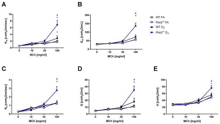
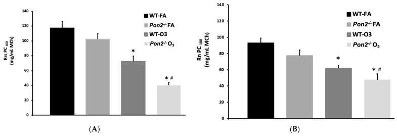
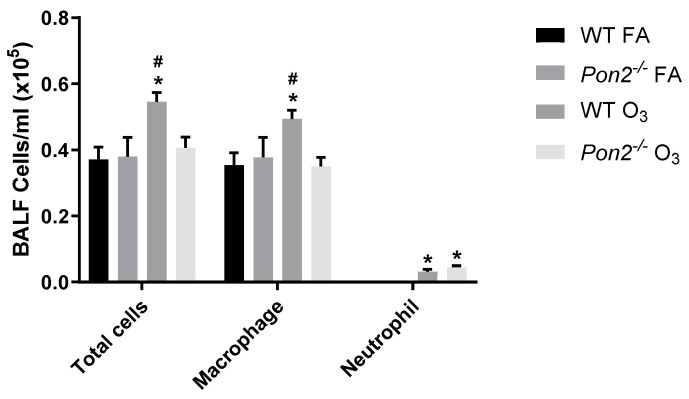
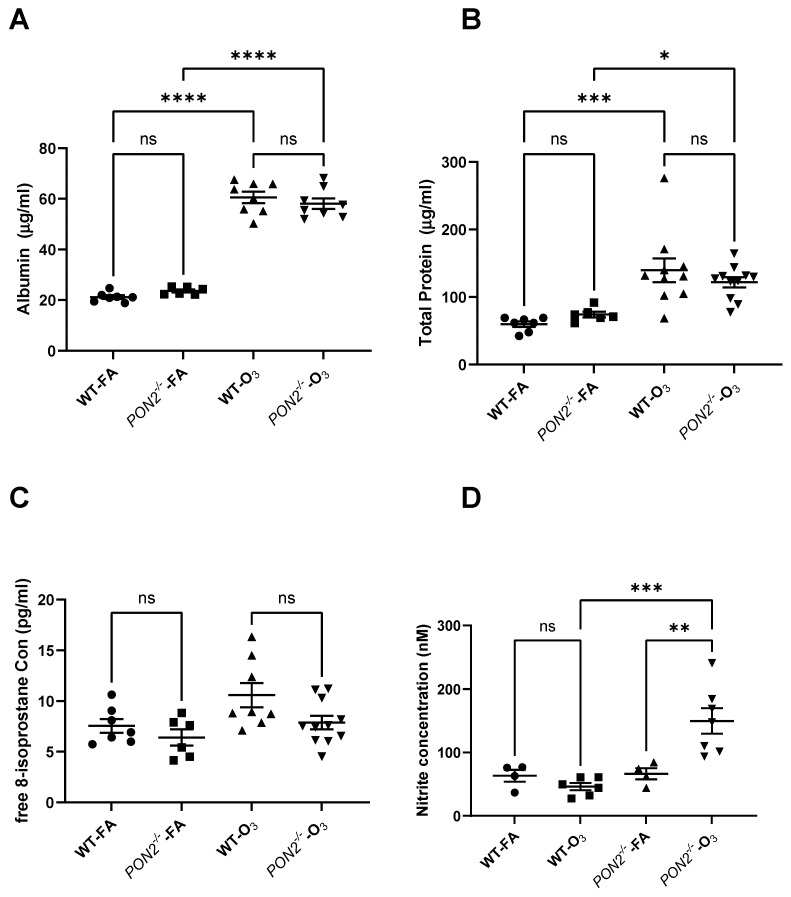
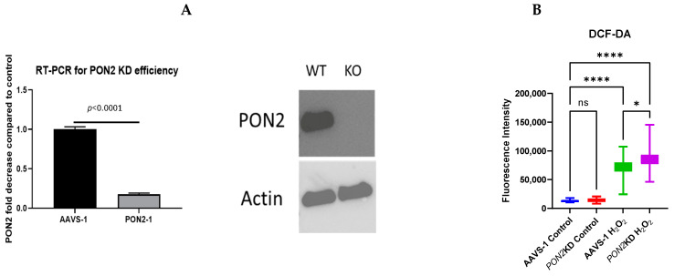
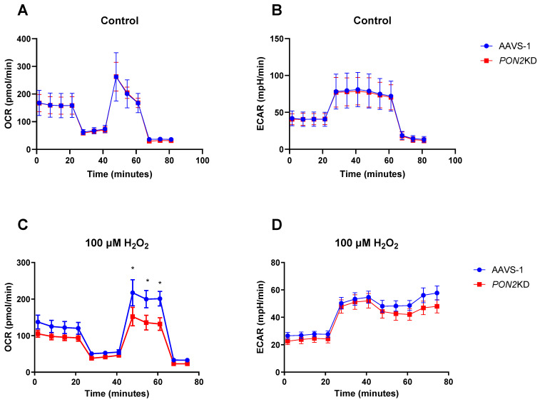
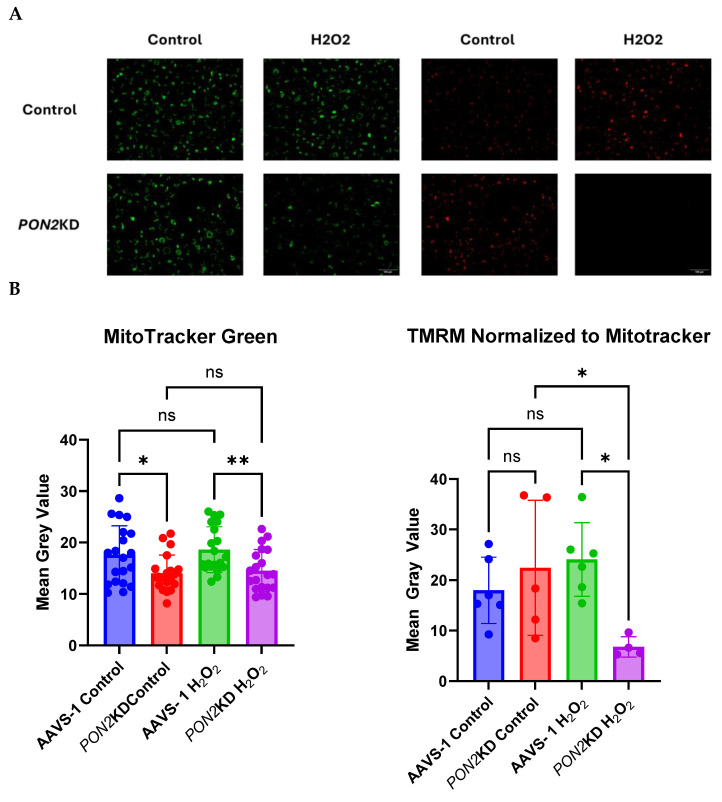
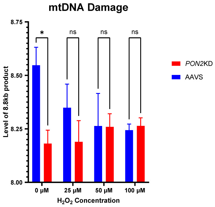
Asthma is a widespread chronic lung disease characterized by airway inflammation and hyperresponsiveness. This airway inflammation is classified by either the presence (T2-high) or absence (T2-low) of high levels of eosinophils. Because most therapies for asthma target eosinophils and related pathways, treatment options for T2-low disease are limited. New pathophysiologic targets are needed. Oxidant stress is a common feature of T2-low disease. Airway epithelial expression of the antioxidant enzyme Paraoxonase 2 (PON2) is decreased in a well-recognized population of people with T2-low asthma and people with obesity and asthma. As a potential mechanism of increased oxidant stress, we measured the role of PON2 in lung oxidant responses using an environmentally relevant in vivo murine oxidant exposure (i.e., ozone) and in vitro studies with an immortalized human airway epithelial cell line BEAS-2B. Pon2-deficient (Pon2-/-) mice developed increased airway hyper-responsiveness compared to wild-type controls. Despite reduced alveolar macrophage influx, Pon2-/- mice exhibited increased nitrite production. In human airway epithelial cells incubated with hydrogen peroxide, PON2 knockdown (PON2KD) decreased mitochondrial function and inner mitochondrial membrane potential. These findings suggest that PON2 functions in defending against airway epithelial oxidant stress. Further studies are needed to elucidate the mechanisms linking PON2, oxidant stress, and asthma pathogenesis.

Keywords: asthma; mitochondria; oxidant stress; ozone; paraoxonase.


Conflict of interest statement

The authors declare no conflicts of interest.

Figures

Figure 1
Figure 1
Airway mechanics measured in wildtype (WT) and Pon2−/− mice 24 h after a 3 h exposure to filtered air (FA) or 2 ppb ozone (O3). Airway responsiveness was determined by administering 0–100 mg/mL aerosolized acetyl-β-methylcholine (methacholine [MCh]). N = 12 for WT FA; N = 5 for Pon2−/− FA; N = 13 for WT O3; and N = 9 for Pon2−/− O3. (A) Rrs, airway resistance; (B) Ers, airway elastance; (C) Rn, Newtonian resistance; (D) G, tissue dampening; and (E) H, tissue elastance. Data presented as mean ± standard deviation. * Pon2−/− vs. FA control, p < 0.05. # Pon2−/− vs. WT, p < 0.05.
Figure 2
Figure 2
(A). PC100 Rrs Provocative concentration of methacholine (MCh) (PC100) that causes a doubling in Rrs from baseline (PC100). FA filtered air; O3 ozone; * p < 0.05 from FA; # p < 0.05 WT- O3 vs. Pon2−/− O3; one-way ANOVA with post-hoc t-test with Welch’s correction factor. (B). PC100 Rn. Provocative concentration of methacholine (MCh) (PC100) that causes a doubling in Rn from baseline (PC100). Data presented as mean +/− standard deviation FA filtered air; O3 ozone; * p < 0.05 from FA; # p < 0.05 WT-O3 vs. Pon2−/− O3 Kruskal–Wallis with post-hoc t-test with Mann–Whitney. N = 12 for WT FA; N = 5 for Pon2−/− FA; N = 13 for WT O3; and N = 9 for Pon2−/− O3.
Figure 3
Figure 3
Total and differential cell counts in BAL fluid measured in Pon2−/− and WT mice 24 h following exposure to filtered air (FA) or 2 ppb ozone (O3). Cell counts were measured using a K2 Cellometer. Differential cell counts were performed by manual count on a single 40x image of a cytospin slide. N = 13 for WT FA; N = 5 for Pon2−/− FA; N = 18 for WT O3; and N = 12 for Pon2−/− O3. Data presented as mean ± standard deviation. Comparison made by 2-way ANOVA. * p < 0.05 compared to FA. # p < 0.05 compared to the other genotype.
Figure 4
Figure 4
Airway permeability and oxidant stress markers in BAL fluid measured in Pon2−/− and WT mice 24 h following exposure to filtered air (FA) or 2 ppb ozone (O3). (AC) Protein, albumin, and 8-isoprostane measured by ELISA. (D) Nitrite was measured by a fluorometric assay. N = 7 for WT FA; N = 6 for Pon2−/− FA; N = 10 for WT O3; and N = 11 for Pon2−/− O3 in the protein and albumin assays. N = 7 for WT FA; N = 6 for Pon2−/− FA; N = 8 for WT O3; and N = 11 for Pon2−/− O3 in the 8-isoprostane assay. N = 4 for WT FA; N = 6 for Pon2−/− FA; N = 4 for WT O3; and N = 7 for Pon2−/− O3 in the nitrite assay. Numbers differ due to availability of BAL fluid sample. Data presented as mean ± standard deviation. Comparison made by 2-way ANOVA. * = p < 0.05, ** = p < 0.01, *** = p < 0.001, **** = p < 0.0001, ns = not significant.
Figure 5
Figure 5
(A). CRISPR PON2 knockdown. PON2 was knocked down in BEAS-2b cells using CRISPR-Cas-9 with AAVS delivery (PON2-1). AAVS-1 cells were exposed to AAVS control. RT-PCR performed by normalizing to expression of the glyceraldehyde-3-phosphate (GAPDH) housekeeping gene. A significant decrease in PON2 gene expression by RT-PCR (left) and PON2 protein expression by western blot (right) was observed. Hypothesis tested by two-tailed t-test. (B). DCF-DA. BEAS-2b cells were plated on a 96 well plate and stained with 200 nM of DCF-DA before being challenged with 100 μM of H2O2 for 4 h. (n = 4/condition). Data presented as mean ± standard deviation. ns = not significant * = p < 0.05, **** = p < 0.0001.
Figure 6
Figure 6
Mitochondrial Function. Cells were exposed to either 100 µM H2O2 or control media for 24 h. 10,000 cells/well were plated in Seahorse XFp plates (n = 3/conditions), and then mitochondrial function was assessed using a MitoStress Test Cartridge. Oxygen Consumption Rate (OCR) is a measure of total electron transport chain function. Extracellular Acidification Rate (ECAR) is a measure of glycolytic flux. (Top Row). No difference was observed in any parameter between the AAVS-1 cells and PON2KD cells under control conditions in both OCR (A) and ECAR (B). (Bottom Row) We observed a reduction in OCR (C) following FCCP addition in PON2KD cells, indicating a decrease in maximal respiration, without changes in baseline respiratory or electron leak. This finding indicates a decrease in mitochondrial space respiratory capacity (SRC). No differences were observed in any ECAR (D) parameter, indicating that changes in OCR were not due to an increase in glycolysis. Comparisons made by 2-way ANOVA with correction for multiple, repeated comparisons. Data presented as mean ± standard deviation * p < 0.05.
Figure 7
Figure 7
Mitochondrial function and mass in AAVS-1 and PON2KD cells. Cells were exposed to 100 µM H2O2 for 24 h. ΔΨm. PON2KD and AAVS-1 control BEAS-2b cells were grown to 70% confluence and stained with (A) MitoTracker Green (Left) and TMRM (Right) and imaged at 40x magnification. (B) Fluorescence intensity of Mitotracker Green (Right) and TMRM normalized to Mitotracker Green (Left). We observed a decrease in total mitochondrial mass in PON2KD cells compared to AAVS-1 control that does not change with H2O2 exposure. However, a decline in relative TMRM expression following H2O2 exposure in PON2KD cells compared to controls was noted, indicating a decrease in ΔΨ. Data presented as mean ± standard deviation. Comparisons made by 2-way ANOVA. ns = not significant, * p < 0.05, ** p < 0.01.
Figure 8
Figure 8
Mitochondrial DNA Lesions. BEAS-2b cells were exposed to 0, 25, 50, or 100 µM of H2O2 for 24 h. mtDNA lesions were measured by amplification of an 8.8 kB fragment and subsequent quantification by RT-PCR. N = 3/group. Data are depicted as means ± standard deviations. Comparisons made with multiple paired t-tests ns = not significant * p < 0.05.

References

    1. Global burden of 369 diseases and injuries in 204 countries and territories, 1990–2019: A systematic analysis for the Global Burden of Disease Study 2019. Lancet. 2020;396:1204–1222. doi: 10.1016/S0140-6736(20)30925-9. - DOI - PMC - PubMed
    1. Woolcock A.J., Salome C.M., Yan K. The Shape of the Dose-Response Curve to Histamine in Asthmatic and Normal Subjects. Am. Rev. Respir. Dis. 1984;130:71–75. doi: 10.1164/arrd.1984.130.1.71. - DOI - PubMed
    1. Kuruvilla M.E., Lee F.E.-H., Lee G.B. Understanding Asthma Phenotypes, Endotypes, and Mechanisms of Disease. Clin. Rev. Allergy Immunol. 2019;56:219–233. doi: 10.1007/s12016-018-8712-1. - DOI - PMC - PubMed
    1. Brusselle G.G., Koppelman G.H. Biologic Therapies for Severe Asthma. N. Engl. J. Med. 2022;386:157–171. doi: 10.1056/NEJMra2032506. - DOI - PubMed
    1. Woo S.-D., Park H.S., Yang E.-M., Ban G.-Y., Park H.-S. 8-Iso-prostaglandin F2α as a biomarker of type 2 low airway inflammation and remodeling in adult asthma. Ann. Allergy Asthma Immunol. 2024;133:73–80.e2. doi: 10.1016/j.anai.2024.04.007. - DOI - PubMed

LinkOut - more resources