. 2024 Nov 5;13(22):1826.
doi: 10.3390/cells13221826.

The Adhesion GPCR ADGRL2/LPHN2 Can Protect Against Cellular and Organismal Dysfunction

Affiliations

The Adhesion GPCR ADGRL2/LPHN2 Can Protect Against Cellular and Organismal Dysfunction

Philipp Jakobs et al. Cells. .

Abstract

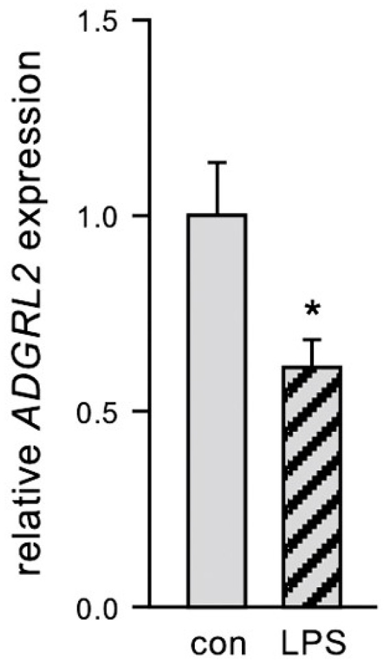
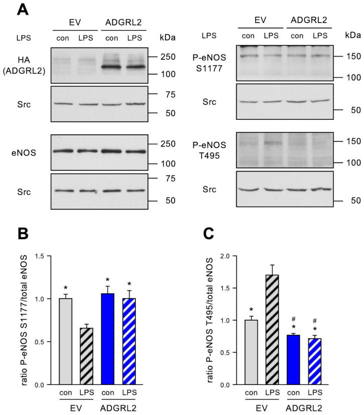
The most common trigger of sepsis and septic shock is bacterial lipopolysaccharide (LPS). Endothelial cells are among the first to encounter LPS directly. Generally, their function is closely linked to active endothelial NO Synthase (eNOS), which is significantly reduced under septic conditions. LPS treatment of endothelial cells leads to their activation and apoptosis, resulting in loss of integrity and vascular leakage, a hallmark of septic shock. Hence, therapies that prevent endothelial leakage or restore the endothelial barrier would be invaluable for patients. Adhesion GPCRs (aGPCRs) have been largely overlooked in this context, although particularly one of them, ADGRL2/LPHN2, has been implicated in endothelial barrier function. Our study shows that overexpression of ADGRL2 protects endothelial cells from LPS-induced activation, apoptosis, and impaired migration. Mechanistically, ADGRL2 preserves eNOS activity by shifting its binding from Caveolin-1 to Heat Shock Protein 90. Furthermore, ADGRL2 enhances antioxidative responses by increasing NRF2 activity. Notably, we found that this function may be evolutionarily conserved. In the absence of lat-2, a homolog of ADGRL2 in Caenorhabditis elegans, worms show higher ROS levels and altered stress response gene expression. Additionally, lat-2 mutants have a significantly reduced lifespan, altogether indicating a protective role of ADGRL2 against oxidative stress across species.

Keywords: ADGRL2/LPHN2; Caenorhabditis elegans; NRF2; eNOS; endothelial cells; lipopolysaccharide; reactive oxygen species; skn-1.


Conflict of interest statement

The authors declare no conflicts of interest.

Figures

Figure 1
Figure 1
LPS downregulates ADGRL2 expression in primary human ECs. ECs were treated for 20 h with 150 ng/mL active LPS (LPS) or detoxified LPS as the control (con). Expression of ADGRL2 was determined by semi-quantitative real-time PCR. RPL32 was used for normalization (data are the mean ± SEM, n = 4, * p < 0.05 vs. con, unpaired, two-sided Student’s t-test).
Figure 2
Figure 2
ADGRL2 prevents LPS-induced activation and apoptosis of ECs and enhances the migratory capacity. ECs were transfected with an expression vector for ADGRL2 or a corresponding empty vector (EV). After transfection, the cells were treated for 20 h with 150 ng/mL active LPS (LPS) or detoxified LPS as the control (con). (A,B) Cells were fixed and stained for ICAM1 (green) and ADGRL2 (red), and nuclei were counterstained with DAPI. (A) Representative immunofluorescence staining (scale bars = 50 μm). (B) Semi-quantitative analysis of ICAM1 levels per cell (data are the mean ± SEM, n = 4, * p < 0.05 vs. EV/LPS, one-way ANOVA with post hoc Tukey LSD test). (C,D) HA-tagged ADGRL2 (HA(ADGRL2)), uncleaved (full-length Caspase-3), and cleaved Caspase-3 were detected by immunoblot, and Src served as the loading control. (C) Representative immunoblots. (D) Semi-quantitative analysis of relative amounts of cleaved Caspase-3 (data are the mean ± SEM, n = 5, * p < 0.05 vs. EV/LPS, one-way ANOVA with post hoc Tukey LSD test). (E,F) The migratory capacity was determined by scratch wound assays. Cell nuclei were stained with DAPI. (E) Representative DAPI staining. Wounds were set at the dashed lines; right of the lines are the cells that had migrated into them (scale bars = 100 µm). (F) Semi-quantitative analysis of migrated cells per high-power field (HPF) (data are the mean ± SEM, n = 5–6, * p < 0.05 vs. EV/LPS, # p < 0.05 vs. EV/con, one-way ANOVA with post hoc Tukey LSD test).
Figure 3
Figure 3
ADGRL2 increases eNOS activity. ECs were transfected with an expression vector for ADGRL2 or a corresponding empty vector (EV). After transfection, the cells were treated for 20 h with 150 ng/mL active LPS (LPS) or detoxified LPS as the control (con). HA-tagged ADGRL2 (HA (ADGRL2)), total eNOS (eNOS), and eNOS phosphorylated at serine 1177 (P-eNOS S1177) or at threonine 495 (P-eNOS T495), respectively, were detected by immunoblot; in all cases, Src served as the loading control. (A) Representative immunoblots; each blot is shown with its respective loading control. (B) Semi-quantitative analysis of the ratio of eNOS phosphorylated at serine 1177 to the total eNOS (data are the mean ± SEM, n = 8, * p < 0.05 vs. EV/LPS, one-way ANOVA with post hoc Tukey LSD test). (C) Semi-quantitative analysis of the ratio of eNOS phosphorylated at threonine 495 to total eNOS (data are the mean ± SEM, n = 5, * p < 0.05 vs. EV/LPS, # p < 0.05 vs. EV/con, one-way ANOVA with post hoc Tukey LSD test).
Figure 4
Figure 4
ADGRL2 alters eNOS interactions with Caveolin-1 and HSP90. ECs were transfected with an expression vector for ADGRL2 or a corresponding empty vector (EV). After transfection, the cells were treated for 20 h with 150 ng/mL active LPS (LPS) or detoxified LPS as the control (con). Interactions between eNOS and Caveolin-1 (CAV1, left panels) and between eNOS and Heat Shock Protein 90 (HSP90, middle panels), respectively, were detected by a proximity ligation assay (green dots). The cytoskeleton was stained with Phalloidin Alexa 594 (red), and nuclei were counterstained with DAPI (blue). Negative controls, in which only one of the primary antibodies was used, are shown in the right panels (scale bars = 50 μm).
Figure 5
Figure 5
ADGRL2 activates endogenous NRF2 and protects against the LPS-induced loss of NRF2. (A) Schematic depiction of luciferase reporter vectors. In all plasmids, expression of firefly luciferase is driven by a minimal promoter derived from the herpes simplex virus thymidine kinase gene containing only a TATA box. The plasmid p(ARE)2TATA-LUC contains two copies of a 41 bl long antioxidant response element (ARE) from the murine Glutathione S-transferase alpha 1 gene promoter directly upstream of the minimal promoter. In p(AREmut)2TATA-LUC, the core sequence of these AREs is mutated to prevent the binding of NRF2. (B) Intact and mutated ARE. Shown is the sequence logo of the core binding sequence of human NRF2 and the corresponding sequence in the ARE of p(ARE)2TATA-LUC (AREwt); the mutated bases in p(AREmut)2TATA-LUC are shown in red (AREmut). (C,D) ECs were co-transfected with the reporter vector containing intact (wt), mutated (mut), or no AREs (–) and an expression vector for ADGRL2 or a or a corresponding empty vector (EV). After transfection, the cells were treated for 20 h with active LPS (LPS) or detoxified LPS as the control (con). (C) Luciferase activity was measured in cell lysates and is shown relative to AREwt/EV/con, which reflects the basal endogenous NRF2 activity (data are the mean ± SEM, n = 9–21, * p < 0.05 vs. AREwt/EV/con, # p < 0.05 vs. AREwt/EV/LPS, n.s. = not significant, one-way ANOVA with post hoc Tukey LSD test). (D) Expression of HA-tagged ADGRL2 (HA (ADGRL2)) in the co-transfections with p(ARE)2TATA-LUC was confirmed by immunoblot, and Src served as the loading control.
Figure 6
Figure 6
Nematodes lacking the latrophilin homolog lat-2 show increased ROS levels, altered expression of stress-related genes, and reduced lifespans. (A) The ROS levels are increased in lat-2(knu505) individuals, both under normal conditions and after exposure to juglone. Worms were lysed after a 1-h treatment with 100 µM juglone (jug), incubated with H2DCF-DA, and DCF conversion was measured over a 2-h period. Control samples (con) were treated with ethanol instead of juglone (data are the mean ± SEM, n = 3, with 5 replicates per n, * p < 0.05 vs. wild-type/con, # p < 0.05 vs. wt/jug, two-way ANOVA with post hoc Bonferroni test for comparing multiple groups). (B) Expression of stress-related genes in lat-2(knu505) hermaphrodites. Expression of single genes was determined by semi-quantitative real-time PCR and normalized to the geometric mean of the reference genes act-1, cdc-42, pmp-3, eif-3.c, and tba-1 and is shown relative to wild-type animals (data are the mean ± SEM, n = 3, with 3 replicates per n, * p < 0.05 vs. wt, unpaired, two-sided Student’s t-test. (C) lat-2(knu505) nematodes have a significantly reduced lifespan (Cox–Mantel test, p < 0.0001). The median survival is 16 days for lat-2(knu505) hermaphrodites and 23 days for wild-type individuals (p < 0.001). n ≥ 200 in 4 independent experiments.

References

    1. Rudd K.E., Johnson S.C., Agesa K.M., Shackelford K.A., Tsoi D., Kievlan D.R., Colombara D.V., Ikuta K.S., Kissoon N., Finfer S., et al. Global, regional, and national sepsis incidence and mortality, 1990–2017: Analysis for the Global Burden of Disease Study. Lancet. 2020;395:200–211. doi: 10.1016/S0140-6736(19)32989-7. - DOI - PMC - PubMed
    1. Merk D., Cox F.F., Jakobs P., Promel S., Altschmied J., Haendeler J. Dose-Dependent Effects of Lipopolysaccharide on the Endothelium-Sepsis versus Metabolic Endotoxemia-Induced Cellular Senescence. Antioxidants. 2024;13:443. doi: 10.3390/antiox13040443. - DOI - PMC - PubMed
    1. Boutagy N.E., McMillan R.P., Frisard M.I., Hulver M.W. Metabolic endotoxemia with obesity: Is it real and is it relevant? Biochimie. 2016;124:11–20. doi: 10.1016/j.biochi.2015.06.020. - DOI - PMC - PubMed
    1. Shu Y., Jin S. Caveolin-1 in endothelial cells: A potential therapeutic target for atherosclerosis. Heliyon. 2023;9:e18653. doi: 10.1016/j.heliyon.2023.e18653. - DOI - PMC - PubMed
    1. Hamann J., Aust G., Arac D., Engel F.B., Formstone C., Fredriksson R., Hall R.A., Harty B.L., Kirchhoff C., Knapp B., et al. International Union of Basic and Clinical Pharmacology. XCIV. Adhesion G protein-coupled receptors. Pharmacol. Rev. 2015;67:338–367. doi: 10.1124/pr.114.009647. - DOI - PMC - PubMed

Publication types

MeSH terms

LinkOut - more resources