. 2024 Nov 6;13(22):1837.
doi: 10.3390/cells13221837.

Short-Term Culture of Human Hyalocytes Retains Their Initial Phenotype and Displays Their Contraction Abilities

Affiliations

Short-Term Culture of Human Hyalocytes Retains Their Initial Phenotype and Displays Their Contraction Abilities

Alessandra Micera et al. Cells. .

Abstract

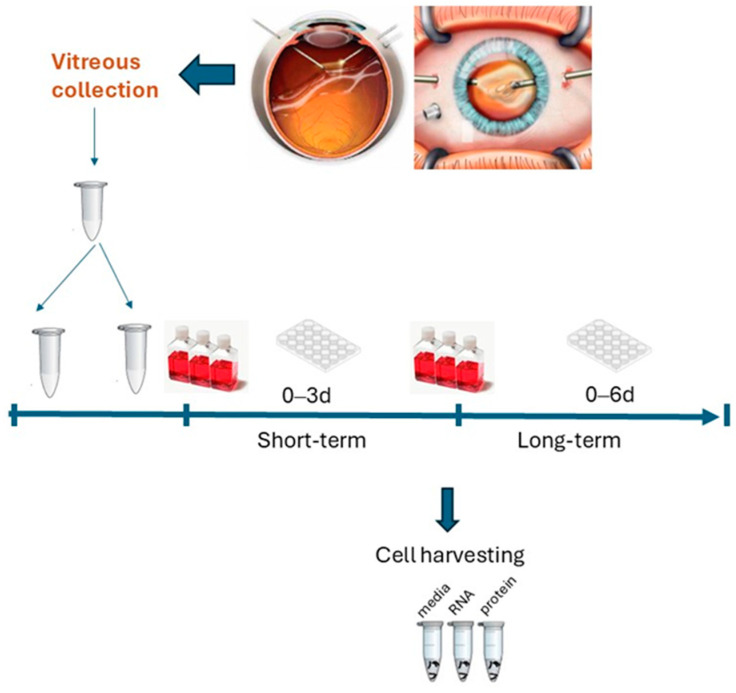
Background: Hyalocytes are the main vitreal cell types with critical functions in health and vitreoretinal diseases. Our aim was to develop cultures of human hyalocytes and verify the retention of their initial cellular features after 3 and 6 days of culturing (3 d and 6 d) by analyzing and comparing a few morphological and functional parameters.

Methods: Vitreous samples (n = 22) were collected and vitreous cells and bead-enriched hyalocytes were developed and compared (3 d vs. 6 d cultures). Vitreous and conditioned media were tested for collagen, vascular endothelial growth factor (VEGF), transforming growth factor β1 (TGFβ1), nerve growth factor (NGF), matrix metalloproteinases (MMPs)/tissue inhibitors of metalloproteinases (TIMPs) and alpha-smooth muscle actin (αSMA) expression (ELISA, array/IP/WB, RT-PCR). Cells were observed at light and fluorescent microscopy (phenotypical properties) and tested for their 3D collagen gel contraction abilities.

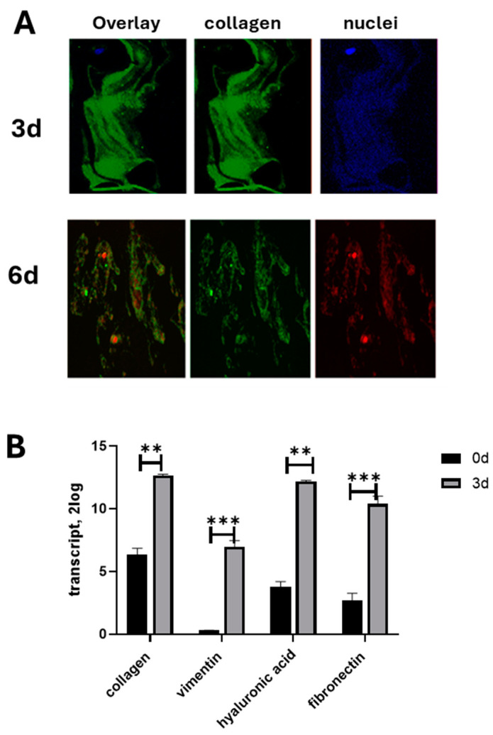
Results: An increased expression of collagens, vimentin, fibronectin, and the MMP9/TIMP1 ratio were observed in vitreous tissues. In 3 d cultures, collagens and MMP9 were upregulated while the related tissue-enzymes were deregulated. Vitreous samples also showed high levels of TGFβ1, VEGF, and NGF, and this protein signature was retained at 3 d while decreased at 6 d. The original phenotype (low αSMA) was retained at 3 d from seeding while an increased αSMA expression was observed at 6 d; NGF/trkANGFR was expressed in cultured hyalocytes and partially drives the collagen retraction.

Conclusions: The vitreous print comparison between untouched and cultured hyalocytes allowed us, on one side, to select 3 d cultures and, on the other, to highlight the neuroprotective/contractile NGF in vitro hyalocytes effects. The possibility of scoring reactive hyalocytes would represent an interesting aspect of screening the vitreoretinal interface severity.

Keywords: NGF; hyalocytes; vitreal cells; vitreoretinal diseases; vitreous; αSMA.


Conflict of interest statement

The authors declare no conflicts of interest. The funders had no role in the design of the study; in the collection, analyses, or interpretation of data; in the writing of the manuscript; or in the decision to publish the results.

Figures

Figure 1
Figure 1
Flow chart of the study. Hyalocytes were extracted from vitreous samples collected at the time of vitreoretinal surgery. Enrichment and bead-based selection were carried out to favor the growth of hyalocytes in a media resembling those of a vitreal chamber (1:1 vitreous-DMEM) shifted after 3 d into complete IMDM. Cultures were directly used for light microscopy or harvested for RNA and protein analysis. Conditioned media from 6 d, 3 d, and 0 d were compared. Adherent cells were quickly fixed in 2% buffered PFA while single cells were directly extracted in lysis solution and devoted to RNA/protein extraction. Conventional detachment with trypsin-EDTA was not used to avoid cell loss at pre-washing and centrifugation.
Figure 2
Figure 2
Human hyalocytes in 3 d and 6 d cultures. Phase contrast microscopy of monolayers acquired from vitreous cells (AC) and hyalocytes (DF) cultured and analyzed after 3 d and 6 d from seeding (0 d). In the light microscope acquisitions, no stains nor dyes were used for evaluation (AC). Note the different cell densities depending on the characteristics of the specimens (AF). (×200, original magnification) Untouched cells displayed different markers of macrophage lineage (G) as detected by PCR analysis. A selected transcript analysis of vitreous cells displays a typical pattern of differentiation (G). The cell survival profile of enriched vitreous cells (H) showed an increased cell number at 3 d and a significant reduction at 6 d (I) with respect to 0 d. The decrease between 6 d and 3 d was significant (p < 0.05; ns not significant, ANOVA Tukey–Kramer post hoc). Bead-selected hyalocytes showed an increase at 3 d and a decrease at 6 d from culturing, with respect to 0 d (C). * p < 0.01.
Figure 3
Figure 3
Immunophenotypical characterization. Epifluorescent images of hyalocytes double-stained with CD64/dapi (A) or triple-stained with CD64/Hoechst/propidium (B). Cells were seeded (1000 cells/well) in 24 well tissue-culture plates and collected at 3 d and 6 d from initial plating (baseline; 0 d). Adherent cells were postfixed and immunostained. Representative images of bead-separated hyalocyte (CD45/CD11a/CD64) cells cultured on round coverslips and imaged at 3 d (B) and 6 d (C) from initial plating (A). Overlay images (upper) display immunoreactivity at 0 d (A), 3 d (B), and 6 d (C); middle layers show CD64 (cy2/green) immunoreactivity changes and lower panels show nuclear intercalants (Hoechst/dapi/blue). (Original magnification ×400).
Figure 4
Figure 4
Hyalocyte cultures partially retain their original phenotype. Representative epifluorescent images of vitreous cells as double-stained with fluorescent collagen type IX marker and nuclear intercalant (DAPI/3d or PI/6d) at 3 d and 6 d time-culturing (A). Cells were seeded (1000 cells/well) in 24 well tissue-culture plates and postfixed at 3 d and 6 d from initial plating (baseline; 0 d). Note the decreased immunoreactivity of collagen matrix after 6 d of culturing. Single acquisitions for collagen type IX are shown in the middle panels (cy2/green). (Original magnification ×200). Transcript analysis showed a significant upregulation at 3 d from plating (B). Note the transcription expression of collagen type-IV, vimentin, hyaluronic acid, and fibronectin transcripts’ expression as assessed by REST-Anova analysis (**: p < 0.001; ***: p < 0.0001).
Figure 5
Figure 5
Tissue enzymes and growth factors are deregulated in 3 d cultures. Analysis of ECM at plating showed the presence of metalloproteinases’ enzymes (MMPs) and related tissue inhibitors (TIMPs) (A). An increased expression was observed for MMP9 at 3 d of culturing, while a significant decrease was monitored for TIMP1/2 (A). Growth factors analysis showed no significant changes in NGF expression while VEGF, TGFβ1, and the other neurotrophins were deregulated (B). ANOVA Tukey–Kramer post hoc analysis.
Figure 6
Figure 6
NGF and trkANGFR expression as a function of culturing. Cells were seeded (1000 cells/well) in 24 well tissue-culture plates and harvested at 3 d and 6 d from initial plating (baseline; 0 d) for NGF quantification or they were immunostained for trkANGFR examination. (A) NGF protein expression as a function of culturing (0 d, 3 d, and 6 d; ANOVA Tukey–Kramer post hoc). Representative epifluorescent images of 3 d hyalocytes (CD45 and CD11a pos selection) cultured for 3 d and identified by nuclear counterstained with dapi (blue; (B)) and double-immunostained for CD64 (green) and trkANGFR (red). As shown in overlay (B), CD64 was mainly localized at the membrane level (green; (D)) and equally distributed, while the trkANGFR receptor appears in membrane zones (red; (E)), nuclei were stained with dapi (C). This distribution cluster of trkANGFR is more visible in the interactive 3D surface plot compared to the CD64 uniform distribution ((F) vs. (G); ImageJ). The nuclear localization by DNA intercalant (dapi/blue) is suggestive of signal specificity. Original magnification ×400 for Merge and single acquisitions.
Figure 7
Figure 7
Hyalocytes express αSMA phenotype and contract a gel matrix upon NGF stimulation. Cells were embedded in a gel matrix (see MM for details) and their ability to contract the matrix was tested in the presence or absence of NGF as a stimulating factor. (A) Hyalocyte RNAs from 3 d- and 6 d cultures were probed for αSMA. A significant transcription of αSMA was observed at 3 d and quickly reduced at 6 d from plating (0 d). (BD) Vitreous cells were drawn off and embedded in a 3D gel contraction pre-stained model. Untreated (black-line) and NGF-supplemented (red-line) vitreous cells were monitored daily for 6 d. Diameters were daily measured, and the results are plotted in (B). Gel retraction occurred by 3 d culturing and was absent at 6 d of culturing. As expected, the range of contraction was strictly related to the αSMA transcription. Representative images from 24 well-plates have 3 d cell-driven retracted gels (B).

Similar articles

Cited by

References

    1. Ribeiro L., Oliveira J., Kuroiwa D., Kolko M., Fernandes R., Junior O., Moraes N., Vasconcelos H., Oliveira T., Maia M. Advances in Vitreoretinal Surgery. J. Clin. Med. 2022;11:6428. doi: 10.3390/jcm11216428. - DOI - PMC - PubMed
    1. Boneva S.K., Wolf J., Hajdú R.I., Prinz G., Salié H., Schlecht A., Killmer S., Laich Y., Faatz H., Lommatzsch A., et al. In-Depth Molecular Characterization of Neovascular Membranes Suggests a Role for Hyalocyte-to-Myofibroblast Transdifferentiation in Proliferative Diabetic Retinopathy. Front. Immunol. 2021;12:757607. doi: 10.3389/fimmu.2021.757607. - DOI - PMC - PubMed
    1. Jones C.H., Gui W., Schumann R.G., Boneva S., Lange C.A., van Overdam K., Chui T.Y., Rosen R.B., Engelbert M., Sebag J. Hyalocytes in proliferative vitreo-retinal diseases. Expert Rev. Ophthalmol. 2022;17:263–280. doi: 10.1080/17469899.2022.2100764. - DOI - PMC - PubMed
    1. Cacciamani A., Parravano M., Scarinci F., Esposito G., Varano M., Micera A. A Simple Spontaneous Vitreal Reflux Collecting Procedure During Intravitreal Injection: Set-Up and Validation Studies. Curr. Eye Res. 2016;41:971–976. doi: 10.3109/02713683.2015.1080282. - DOI - PubMed
    1. Cacciamani A., Esposito G., Scarinci F., Parravano M., Dinice L., Di Nicola M., Micera A. Inflammatory mediators in the vitreal reflux of patients with diabetic macular edema. Graefe’s Arch. Clin. Exp. Ophthalmol. 2019;257:187–197. doi: 10.1007/s00417-018-4169-4. - DOI - PubMed

LinkOut - more resources