. 2024 Nov 7;13(22):1844.
doi: 10.3390/cells13221844.

Modelling Functional Thyroid Follicular Structures Using P19 Embryonal Carcinoma Cells

Affiliations

Modelling Functional Thyroid Follicular Structures Using P19 Embryonal Carcinoma Cells

Fatimah Najjar et al. Cells. .

Abstract

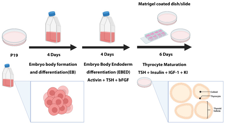
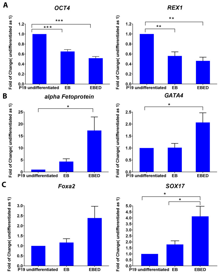
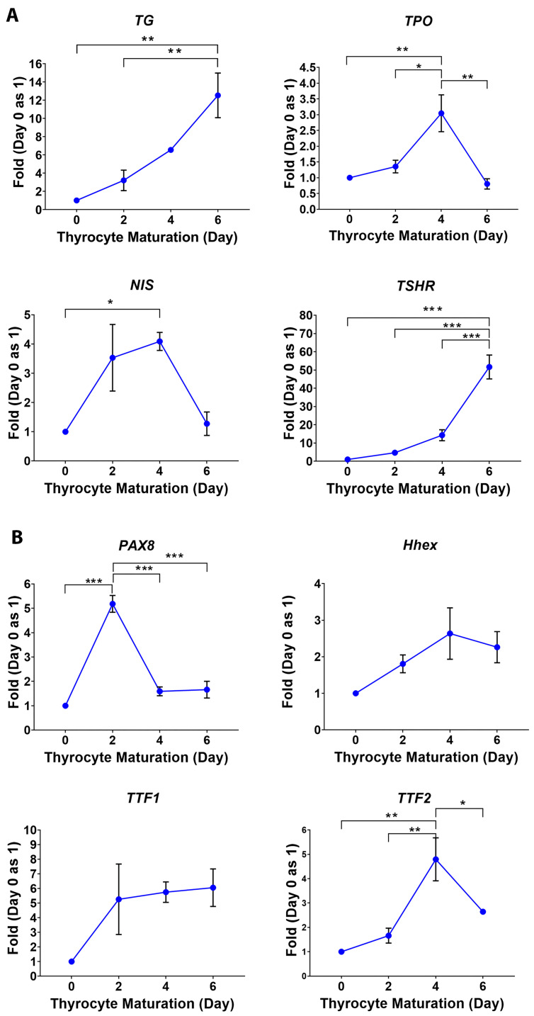
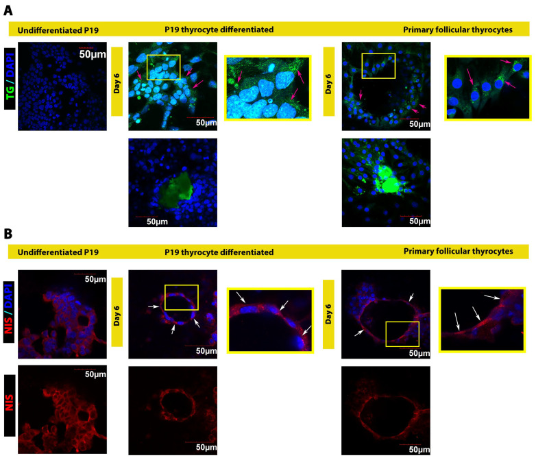
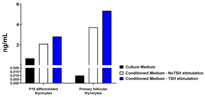
Thyroid gland diseases remain clinical challenges due to the lack of reliable in vitro models to examine molecular pathways of thyrocytes development, maturation, and functional maintenance. This study aimed to develop in vitro thyrocytes model using a stem cell culture, P19 embryonal carcinoma which requires no feeder layer, differentiation into mature and functional thyrocytes that allow molecular and genetic manipulation for studying thyroid diseases. The procedure utilizes Activin A and thyroid stimulating hormone (TSH) to first induce embryoid body endoderm formation enriched in thyrocyte progenitors. Following dissociating embryoid bodies, thyrocyte progenitors are plated in Matrigel as monolayer cultures that allows thyrocyte progenitors mature to functional thyrocytes. These thyrocytes further maturate to form follicle-like structures expressing and accumulating thyroglobulin that can be secreted into the medium upon TSH stimulation. Thyrocyte differentiation-maturation process is monitored by the expression of essential transcriptional factors and thyrocyte-specific functional genes. Further, the applicability of this system is validated by introducing a siRNA control. Following molecular manipulation, the system can still be guided to differentiate into mature and functional thyrocytes. This system spans a time frame of 14 days, suitable for detailed molecular studies to dissect pathways and molecular players in thyrocytes development and functional maintenance.

Keywords: cell differentiation; embryonal carcinoma stem cells; in vitro models; thyrocytes; thyroid diseases.


Conflict of interest statement

The authors declare no conflicts of interest.

Figures

Figure 1
Figure 1
Schematic procedure of P19 differentiation into thyrocytes. Undifferentiated P19 cells are seeded for four days to form embryoid bodies (EBs), followed by four days of incubation with a supplemented medium to differentiate into EB endoderm differentiation (EBED) containing thyrocyte progenitors. The aggregates of EBED are dissociated into single cells and seeded on a Matrigel-coated dish, where cells matured to form follicle-like structures as shown in the bottom of this figure.
Figure 2
Figure 2
Gene expression patterns at different stages of P19 transition from the undifferentiated state to EB endoderm differentiation (EBED) stage. (A) Expression of stemness genes: OCT4 and REX1 in undifferentiated P19 cells, EBs, and EBED. (B) Expression of endoderm-specific genes: α-fetoprotein and GATA4 in undifferentiated P19 cells, EB, and EBED. (C) Expression of genes specific to thyrocyte progenitors and definitive EBED: Foxa2 and SOX17 in undifferentiated P19 cells, EB, and EBED. The data show means ± SE of 3 independent experiments. One-way ANOVA followed by Bon-ferroni post hoc analysis was performed, * p <0.05, ** p < 0.01, *** p < 0.001.
Figure 3
Figure 3
Gene expression patterns of P19 differentiated thyrocytes during maturation. (A) Expression of genes specific to thyrocyte functions: TG and TPO, and those enriched in thyrocytes NIS and TSHR. (B) Expression of transcription factors: Pax8, TTF1, TTF2 and Hhex. The data show means ± SE of 3 independent experiments. One-way ANOVA followed by Bon-ferroni post hoc analysis was performed, * p <0.05, ** p < 0.01, *** p < 0.001.
Figure 4
Figure 4
Immunostaining of TG and NIS protein in undifferentiated P19, P19 differentiated thyrocytes and primary follicular thyrocytes. (A) Upper panel: intracellular TG protein expression (TG, green stain) in undifferentiated P19, P19 differentiated thyrocytes and primary follicular thyrocytes. The intracellular TG was distributed evenly throughout thyrocytes, indicated by magenta arrows. Lower panel: TG staining in the colloid of follicle-like-structures formed by P19 differentiated thyrocytes and primary follicular thyrocytes. The heatmaps/histograms were obtained, shown in Figure S1. (B) Upper panel: NIS expression (NIS, red stain) in undifferentiated P19, P19 differentiated thyrocytes, and primary follicular thyrocytes. NIS was detected mainly on one side (red stain) of the cells (nuclei stained with DAPI), indicating the polarization of thyrocytes (white arrows). The lower panel showed the polarization of NIS pattern.
Figure 5
Figure 5
TSH stimulates TG secretion. TG secreted from P19-differentiated thyrocytes and primary follicular thyrocytes was detected by ELISA. Condition media were collected from three sets of experiments with two days of TSH stimulation. Culture medium was included as the control in each experiment. Primary follicular thyrocyte culture was stimulated with a physiological dose of TSH (1 mIU/mL); whereas P19-thyrocyte culture readily contained a basal level TSH (1 mIU/mL), therefore a higher dose of TSH (10 mIU/mL) was used to stimulate TG secretion.
Figure 6
Figure 6
Gene expression patterns at different stages of P19-siRNA control culture transitioning from undifferentiated state to EB endoderm differentiation (EBED) stage. (A) Expression of stemness genes OCT4 and REX1 in P19-siRNA control, including stages of undifferentiated, EBs, and EBED. (B) Expression of endoderm-specific genes: α-fetoprotein and GATA4 in P19-siRNA control, including stages of undifferentiated, EB, and EBED. (C) Expression of genes specific to thyrocyte progenitors and definitive EBED: Foxa2 and SOX17 in P19-siRNA control, including undifferentiated, EB, and EBED. The data show means ± SE of 3 independent experiments. One-way ANOVA followed by Bonferroni post hoc analysis was performed, * p <0.05, ** p < 0.01, *** p < 0.001.
Figure 7
Figure 7
Gene expression patterns and immunostaining of P19-siRNA control differentiated into thyrocytes. (A) Expression patterns of genes specific to thyrocyte functions: TG and TPO, and those enriched in thyrocytes, NIS and TSHR in P19-wild type and P19-siRNA control cultures thyrocytes differentiated. (B) Immunostaining of TG and NIS in P19-siRNA control cells-differentiated follicle-like structures. Left panel, intracellular TG was distributed evenly in P19-siRNA control cells-differentiated thyrocytes (magenta arrows), and secreted TG accumulated inside the follicle (middle panel). Right panel, NIS was detected in P19-siRNA control cells-differentiated thyrocytes only on one side of the cells, indicating the expected polarity of these differentiated thyrocytes (white arrows). The heatmaps/histograms of TG and nuclear staining were obtained, shown in Figure S2. Panel A data show means ± SE of 3 independent experiments. Two-way ANOVA followed by Bonferroni post hoc analysis was performed, * p <0.05, ** p < 0.01.

Similar articles

Cited by

References

    1. Toda S., Koike N., Sugihara H. Thyrocyte integration, and thyroid folliculogenesis and tissue regeneration: Perspective for thyroid tissue engineering. Pathol. Int. 2001;51:403–417. doi: 10.1046/j.1440-1827.2001.01218.x. - DOI - PubMed
    1. Toda S., Aoki S., Uchihashi K., Matsunobu A., Yamamoto M., Ootani A., Yamasaki F., Koike E., Sugihara H. Culture models for studying thyroid biology and disorders. ISRN Endocrinol. 2011;2011:275782. doi: 10.5402/2011/275782. - DOI - PMC - PubMed
    1. Tomás G., Tarabichi M., Gacquer D., Hébrant A., Dom G., Dumont J.E., Keutgen X., Fahey T.J., Maenhaut C., Detours V. A general method to derive robust organ-specific gene expression-based differentiation indices: Application to thyroid cancer diagnostic. Oncogene. 2012;31:4490–4498. doi: 10.1038/onc.2011.626. - DOI - PubMed
    1. Najjar F., Nhieu J., Wei C.-W., Milbauer L., Burmeister L., Seelig D., Wei L.-N. Deleting Cellular Retinoic-Acid-Binding Protein-1 (Crabp1) Gene Causes Adult-Onset Primary Hypothyroidism in Mice. Endocrines. 2023;4:138–150. doi: 10.3390/endocrines4010013. - DOI
    1. Nilsson M., Fagman H. Development of the thyroid gland. Development. 2017;144:2123–2140. doi: 10.1242/dev.145615. - DOI - PubMed

Publication types

LinkOut - more resources