. 2024 Nov 8;13(22):1857.
doi: 10.3390/cells13221857.

Semaglutide Ameliorates Diabetic Neuropathic Pain by Inhibiting Neuroinflammation in the Spinal Cord

Affiliations

Semaglutide Ameliorates Diabetic Neuropathic Pain by Inhibiting Neuroinflammation in the Spinal Cord

Sing-Ong Lee et al. Cells. .

Abstract

Glucagon-like peptide 1 (GLP-1) receptor agonists are frequently used to treat type 2 diabetes and obesity. Despite the development of several drugs for neuropathic pain management, their poor efficacy, tolerance, addiction potential, and side effects limit their usage. Teneligliptin, a DPP-4 inhibitor, has been shown to reduce spinal astrocyte activation and neuropathic pain caused by partial sciatic nerve transection. Additionally, we showed its capacity to improve the analgesic effects of morphine and reduce analgesic tolerance. Recent studies indicate that GLP-1 synthesized in the brain activates GLP-1 receptor signaling pathways, essential for neuroprotection and anti-inflammatory effects. Multiple in vitro and in vivo studies using preclinical models of neurodegenerative disorders have shown the anti-inflammatory properties associated with glucagon-like peptide-1 receptor (GLP-1R) activation. This study aimed to investigate the mechanism of antinociception and the effects of the GLP-1 agonist semaglutide (SEMA) on diabetic neuropathic pain in diabetic rats.

Methods: Male Wistar rats, each weighing between 300 and 350 g, were categorized into four groups: one non-diabetic sham group and three diabetic groups. The diabetic group received a single intraperitoneal injection of streptozotocin (STZ) at a dosage of 60 mg/kg to induce diabetic neuropathy. After 4 weeks of STZ injection, one diabetic group was given saline (vehicle), and the other two were treated with either 1× SEMA (1.44 mg/kg, orally) or 2× SEMA (2.88 mg/kg, orally). Following a 4-week course of oral drug treatment, behavioral, biochemical, and immunohistochemical analyses were carried out. The mechanical allodynia, thermal hyperalgesia, blood glucose, advanced glycation end products (AGEs), plasma HbA1C, and spinal inflammatory markers were evaluated.

Results: SEMA treatment significantly reduced both allodynia and hyperalgesia in the diabetic group. SEMA therapy had a limited impact on body weight restoration and blood glucose reduction. In diabetic rats, SEMA lowered the amounts of pro-inflammatory cytokines in the spinal cord and dorsal horn. It also lowered the activation of microglia and astrocytes in the dorsal horn. SEMA significantly reduced HbA1c and AGE levels in diabetic rats compared to the sham control group.

Conclusions: These results indicate SEMA's neuroprotective benefits against diabetic neuropathic pain, most likely by reducing inflammation and oxidative stress by inhibiting astrocyte and microglial activity. Our findings suggest that we can repurpose GLP-1 agonists as potent anti-hyperalgesic and anti-inflammatory drugs to treat neuropathic pain without serious side effects.

Keywords: GLP-1RA; SEMA; astrocytes; diabetic neuropathic pain; microglia; pro-inflammatory cytokines.


Conflict of interest statement

The authors declare no conflicts of interest. The funders had no role in the design of the study; in the collection, analyses, or interpretation of data; in the writing of the manuscript; or in the decision to publish the results.

Figures

Figure 1
Figure 1
Overview of the experimental design.
Figure 2
Figure 2
(a) Rats’ mean mechanical paw withdrawal threshold and (b) mean thermal paw withdrawal latency were normalized following a single intraperitoneal injection of streptozotocin (STZ, 60 mg/kg i.p.). The withdrawal threshold of the rats’ paws and thermal latency were assessed each week post injection (n = 6). Control rats received the same volume of normal saline (n = 6) 4 weeks after STZ injection. From week 4, SEMA was orally administered at a dose of 1× (1.44 mg/kg) or 2× (2.88 mg/kg) to rats daily for 4 weeks (week 4–8). Data are presented as mean ± SEM (n = 6). a p < 0.05 vs. Sham; b p < 0.05 vs. DNP group.
Figure 3
Figure 3
(a) Blood glucose concentrations (mg/dL) (b) Changes in body weight and (c) Hb1Ac levels measured at different weeks after a single injection of streptozotocin (60 mg/kg i.p). Sham rats received the same volume of normal saline (n = 6). 4 weeks after STZ injection, SEMA administered at a dose of 1× (1.44 mg/kg) or 2× (2.88 mg/kg) to rats daily for 4 weeks (week 4–8). Data are presented as mean ± SEM (n = 6). a p < 0.05 vs. Sham; b p < 0.05 vs. STZ-DNP group; c p < 0.05 vs. DNP + SEMA 1×.
Figure 4
Figure 4
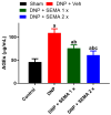
The effect of STZ-induced diabetes on plasma levels of advanced glycation end products (AGEs) in rats with STZ-induced diabetic neuropathy, assessed four weeks after SEMA administration. Data are presented as mean ± SEM (n = 6). a p < 0.05 vs. Sham; b p < 0.05 vs. STZ-DNP group; c p < 0.05 vs. DNP + SEMA 1×.
Figure 5
Figure 5
SEMA’s effects on the levels of (a) TNF-α, (b) IL-1β, and (c) IL-6, in the spinal dorsal horn of the DNP rats. The spinal dorsal horn’s IL-1β, TNF- α, and IL-6 levels were measured using ELISA. Data are presented as mean ± SEM (n = 6). a p < 0.05 vs. Sham; b p < 0.05 vs. STZ-DNP group; c p < 0.05 vs. DNP + SEMA 1×.
Figure 6
Figure 6
(A) Impact of SEMA treatment on microglial cell expression in the spinal cords of the DNP rats. Four weeks post-drug therapy, spinal cord slices were fixed and labeled with the IBA-1 microglial cell marker, followed by imaging via fluorescence microscopy. The sections were obtained from (a) sham rats, (b) DNP rats (c) DNP + SEMA 1× and (d) DNP + SEMA 2×. (B) The quantitative analysis of activated microglial cells is conducted. The yellow arrows in the enlarged portion of figure (a) indicate microglia displaying a resting ramified phenotype, characterized by a small cell body with fewer than one branching process. The red circles in the magnified region of figure (b) indicate microglia displaying an amoeboid morphology, defined by a prominent cell body and multiple branching processes. An asterisk in figure (B) indicates a statistically significant difference among sham versus DNP + Veh, DNP + Veh versus DNP + SEMA 1.44 mg/kg, and DNP + SEMA 1.44 mg/kg versus DNP + SEMA 2.88 mg/kg * p < 0.05; *** p < 0.001, (n = 6 animals per group).
Figure 7
Figure 7
(A) The effect of SEMA treatment on astroglial GFAP staining in the spinal cords of the DNP rats. Four weeks post-drug therapy, spinal cord slices were preserved, labeled with the GFAP marker, and analyzed via fluorescence microscopy. The sections were obtained from (a) sham rats, (b) DNP rats (c) DNP + SEMA 1× and (d) DNP + SEMA 2×. (B) The quantitative analysis of activated astroglial cells is presented. STZ rats exhibited GFAP-positive cells characterized by the typical stellate morphology of astrocytes, featuring numerous elaborate processes highlighted in red circles. Both 1× and 2× reduced astrogliosis. An asterisk indicates a statistically significant difference among sham versus DNP + Veh, DNP + Veh versus DNP + SEMA 1.44 mg/kg, and DNP + SEMA 1.44 mg/kg versus DNP + SEMA 2.88 mg/kg * p < 0.05; *** p < 0.001, (n = 6 animals per group).
Figure 8
Figure 8
(A) The effect of SEMA treatment on TNF-α expression in the spinal cords of the DNP rats. Four weeks post-drug therapy, spinal cord slices were preserved, labeled with the TNF-α marker, and analyzed via fluorescence microscopy. The sections were obtained from (a) sham rats, (b) DNP rats (c) DNP + SEMA 1× and (d) DNP + SEMA 2×. (B) The quantitative analysis of TNF-α-positive cells. TNF-α is produced by various cell types within the spinal cord. (1) Large pyramidal cells, similar to neurons, are indicated by black arrows; (2) solitary cells resembling lymphocytes are highlighted with red circles; and (3) clusters of inflammatory cells are marked by blue arrows. An asterisk indicates a statistically significant difference among sham versus DNP + Veh, DNP + Veh versus DNP + SEMA 1.44 mg/kg, and DNP + SEMA 1.44 mg/kg versus DNP + SEMA 2.88 mg/kg *** p < 0.001, (n = 6 animals per group).
Figure 9
Figure 9
(A) Impact of SEMA treatment on IL-1β expression in the spinal cords of the DNP rats. Four weeks post-drug therapy, spinal cord slices were preserved and labeled with the IL-1β marker, followed by image acquisition via fluorescence microscopy. The sections were obtained from (a) sham rats, (b) DNP rats (c) DNP + SEMA 1× and (d) DNP + SEMA 2×. (B) The quantitative analysis of IL-1β-positive cells. In the DNP rats, a significant increase in IL-1β levels in the spinal cord was noted in comparison to sham animals. The administration of SEMA 1× resulted in a slight reduction of IL-1β expression, while animals injected with DNP and SEMA 2× exhibited staining comparable to that of sham animals. An asterisk indicates a statistically significant difference among sham versus DNP + Veh, DNP + Veh versus DNP + SEMA 1.44 mg/kg, and DNP + SEMA 1.44 mg/kg versus DNP + SEMA 2.88 mg/kg. ** p < 0.01; *** p < 0.001, (n = 6 animals per group).

References

    1. Murnion B.P. Neuropathic pain: Current definition and review of drug treatment. Aust. Prescr. 2018;41:60–63. doi: 10.18773/austprescr.2018.022. - DOI - PMC - PubMed
    1. Rugnath R., Orzechowicz C., Newell C., Carullo V., Rugnath A. A Literature Review: The Mechanisms and Treatment of Neuropathic Pain—A Brief Discussion. Biomedicines. 2024;12:204. doi: 10.3390/biomedicines12010204. - DOI - PMC - PubMed
    1. Chen Y., Song X.J. Diabetic Neuropathic Pain: Directions for Exploring Treatments. Biomedicines. 2024;12:589. doi: 10.3390/biomedicines12030589. - DOI - PMC - PubMed
    1. Cooper T.E., Chen J., Wiffen P.J., Derry S., Carr D.B., Aldington D., Cole P., Moore R.A. Morphine for chronic neuropathic pain in adults. Cochrane Database Syst. Rev. 2017;5:CD011669. doi: 10.1002/14651858.CD011669.pub2. - DOI - PMC - PubMed
    1. Zhang X., Bao L., Shi T.J., Ju G., Elde R., Hökfelt T. Down-regulation of mu-opioid receptors in rat and monkey dorsal root ganglion neurons and spinal cord after peripheral axotomy. Neuroscience. 1998;82:223–240. doi: 10.1016/S0306-4522(97)00240-6. - DOI - PubMed

Publication types

MeSH terms

LinkOut - more resources