. 2024 Nov 11;13(22):1863.
doi: 10.3390/cells13221863.

Immunohistochemical Characterization of Spermatogenesis in the Ascidian Ciona robusta

Affiliations

Immunohistochemical Characterization of Spermatogenesis in the Ascidian Ciona robusta

Haruka Sakurai et al. Cells. .

Abstract

Animals show diverse processes of gametogenesis in the evolutionary pathway. Here, we characterized the spermatogenic cells in the testis of the marine invertebrate Ciona robusta. Ciona sperm differentiate in a non-cystic type of testis, comprising many follicles with various sizes and stages of spermatogenic cells. In the space among follicles, we observed free cells that were recognized by antibody against Müllerian inhibiting substance, a marker for vertebrate Sertoli cells. We further categorized the spermatogenic cells into four round stages (RI to RIV) and three elongated stages (EI to EIII) by morphological and immunohistochemical criteria. An antibody against a vertebrate Vasa homolog recognized a few large spermatogonium-like cells (RI) near the basal wall of a follicle. Consistent with the period of meiosis, a synaptonemal complex protein SYCP3 was recognized from early spermatocytes (RII) to early spermatids (E1). Acetylated tubulins were detected in spermatids before flagellar elongation at the RIV stage and became distributed along the flagella. Electron microscopy showed that the free cells outside the testicular follicle possessed a characteristic of vertebrate Sertoli cells. These results would provide a basis for basic and comparative studies on the mechanism of spermatogenesis.

Keywords: Sertoli cell; ascidian sperm; cilia and flagella; spermatogenesis.


Conflict of interest statement

The authors declare no conflicts of interest.

Figures

Figure 1
Figure 1
Paraffin section images of testicular follicles in the ascidian Ciona robusta. (A) A section of the testis, showing that it is composed of numerous testicular follicles (yellow circle). (B) Distribution of spermatogenic cells within a testicular follicle. Cells with round nuclei are located in the peripheral region, while mature sperm are seen in the central region, connected to the efferent duct (dashed line). Abbreviation: ef, efferent duct. (C,D) Localization of different stages of spermatogenic cells within a follicle. The areas highlighted by dashed squares in (C) are shown at higher magnification in (D).
Figure 2
Figure 2
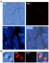
Immunofluorescence image of a frozen section stained with anti-Müllerian Inhibiting Substance (MIS) antibody. (A) MIS H-300 positive cells (red) are localized in the space between testicular follicles. Scale bar, 20 μm. (B) The cells located between follicles display a round shape. Scale bar, 5 μm. Blue, DAPI staining.
Figure 3
Figure 3
Immunofluorescence image of a frozen section stained with an antibody against Ciona robusta Vasa homolog (CiVH). (A) CiVH signals (red) are detected in a few large cells located along the basal region of a testicular follicle. Scale bar, 20 μm. (B) CiVH-positive cells appear round and large, likely representing primordial germ cells or spermatogonia (RI). The immunofluorescence signal was present throughout the cytoplasm but not in the nucleus (blue). Scale bar, 5 μm.
Figure 4
Figure 4
Immunofluorescence image of a frozen section stained with an antibody against TD09. This protein is one of the gene products from highly expressed genes in Ciona testis, showing homology with the synaptonemal complex protein SYCP3. (A) The antibody recognizes a broad area of the testis (red), except for the central region near the efferent duct containing mature sperm. Scale bar, 20 μm. (B,C) The anti-TD09 antibody detects the nuclei of both round-stage cells (RII to RIV; (B)) and short-flagellated elongated-stage cells (EI; (C)). Scale bar, 5 μm. Blue, DAPI staining.
Figure 5
Figure 5
Immunofluorescence image of a frozen section stained with an antibody against TD02. This protein is one of the gene products from highly expressed genes in Ciona testis, showing homology with histone H1-like protein or protamine. (A) The antibody recognizes spermatids and mature sperm (red) located in the central area of the testicular follicle near the efferent duct. Scale bar, 20 μm. (BD) The antibody stained the nuclei of late spermatids and mature sperm (EII; (C), EIII; (D)) but not early-stage spermatids (EI; (B)), even though the latter had already formed long flagella. Scale bar, 5 μm. Red, TD02 immunostaining. Blue, DAPI staining.
Figure 6
Figure 6
Immunofluorescence image of a frozen section stained with anti-acetylated α-tubulin antibody. (A) Immunofluorescence analysis of a testis section showed strong staining in the central area of the testicular follicle, similar to anti-TD02 staining. Scale bar, 20 μm. (B) The antibody recognizes the cytoplasm of non-flagellated spermatids (EI stage). Scale bar, 5 μm. (C) The flagella of mature sperm were strongly stained by the anti-acetylated α-tubulin antibody. Scale bar, 5 μm. Red, immunostaining with anti-acetylated α-tubulin antibody. Blue, DAPI staining.
Figure 7
Figure 7
Transmission electron microscopy images of Ciona testis. (A) An image of a testicular follicle shows the non-cystic distribution of spermatogenic cells. A free cell with prominent heterochromatin in the nucleus (asterisk) is visible between testicular follicles. Scale bar, 5 μm. (B) A higher magnification of the free cell (asterisk). Scale bar, 2 μm. (C,D) Free cells (asterisks) are sometimes associated with the outer layer of a testicular follicle. Scale bars, 2 μm (C); 5 μm (D). (E,F) Long striated rootlets extend from the basal body of a flagellum in flat epithelial cells. Scale bars, 1 μm. (G) The axonemes exhibit a 9+2 structure with dynein arms, indicating motility. Scale bar, 500 nm. Arrows, striated rootlets.
Figure 8
Figure 8
A schematic drawing of Ciona spermatogenesis based on the staining patters by several marker antibodies. Spermatogenic cells are categorized into four round stages (RI to RIV) and three elongated stages (EI to EIII). The reactivities against the antibodies used in this study and the localizations are indicated by colors. Stages at RI, RII/RIII and RIV/EI/EII/EIII are considered as spermatogonia, spermatocytes, and spermatids, respectively (see text).

Similar articles

References

    1. Russell L.D., Ettlin R.A., Hikim A.P., Clegg E.D. Mammalian spermatogenesis. In: Russell L.D., Ettlin R., Sinha Hikim A.P., Clegg E.D., editors. Histological and Histopathological Evaluation of the Testis. Cache River; Clearwater, FL, USA: 1990. pp. 1–40.
    1. Griswold M.D. Spermatogenesis: The Commitment to meiosis. Physiol. Rev. 2016;96:1–17. doi: 10.1152/physrev.00013.2015. - DOI - PMC - PubMed
    1. Yoshida S. From cyst to tubule: Innovations in vertebrate spermatogenesis. Wiley Interdiscip. Rev. Dev. Biol. 2016;5:119–131. doi: 10.1002/wdev.204. - DOI - PMC - PubMed
    1. Satoh N. Developmental Genomics of Ascidians. Wiley-Blackwell; Hoboken, NJ, USA: 2014. pp. 1–216.
    1. Inaba K., Padma P., Satouh Y., Shin-I T., Kohara Y., Satoh N., Satou Y. EST analysis of gene expression in testis of the ascidian Ciona intestinalis. Mol. Reprod. Dev. 2002;62:431–445. doi: 10.1002/mrd.10131. - DOI - PubMed

Publication types

LinkOut - more resources