. 2024 Nov 14;13(22):1882.
doi: 10.3390/cells13221882.

Loss of Dnah5 Downregulates Dync1h1 Expression, Causing Cortical Development Disorders and Congenital Hydrocephalus

Affiliations

Loss of Dnah5 Downregulates Dync1h1 Expression, Causing Cortical Development Disorders and Congenital Hydrocephalus

Koichiro Sakamoto et al. Cells. .

Abstract

Dnah5 is associated with primary ciliary dyskinesia in humans. Dnah5-knockout (Dnah5-/- mice develop acute hydrocephalus shortly after birth owing to impaired ciliary motility and cerebrospinal fluid (CSF) stagnation. In contrast to chronic adult-onset hydrocephalus observed in other models, this rapid ventricular enlargement indicates additional factors beyond CSF stagnation. Herein, we investigated the contributors to rapid ventricular enlargement in congenital hydrocephalus. Dnah5-/- mice were generated using CRISPR/Cas9. The expression of dynein, N-cadherin, and nestin in the cerebral cortex was assessed using microarrays and immunostaining. Real-time PCR and Western blotting were performed for gene and protein quantification, respectively. All Dnah5-/- mice developed hydrocephalus, confirmed by electron microscopy, indicating the absence of axonemal outer dynein arms. Ventricular enlargement occurred rapidly, with a 25% reduction in the number of mature neurons in the motor cortex. Dync1h1 expression was decreased, while cytoplasmic dynein levels were 56.3% lower. Levels of nestin and N-cadherin in the lateral ventricular walls decreased by 31.7% and 33.3%, respectively. Reduced cytoplasmic dynein disrupts neurogenesis and axonal growth and reduces neuron cortical density. Hydrocephalus in Dnah5-/- mice may result from cortical maldevelopment due to cytoplasmic dynein deficiency, further exacerbating ventricular enlargement due to CSF stagnation caused by impaired motile ciliary function.

Keywords: Dnah5; cytoplasmic dynein; motile cilia; neurogenesis; primary cilia.


Conflict of interest statement

The authors declare no conflicts of interest.

Figures
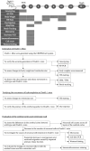
Figure A1
Figure A1
The timeline and flowchart illustrate our research process. These experiments were conducted in three phases. First, knockout mice were generated using the CRISPR/Cas9 system. The accuracy of knockout construction was confirmed through pyrosequencing-based genotyping and RT-PCR. Dnah5−/− mice were then compared with wild-type mice, assessing body weight differences at various postnatal days. HE staining of brain sections was performed, and ventricular size was evaluated in coronal sections. At postnatal day 3, cilia were observed using SEM and TEM. At postnatal day 10, microbeads were applied to coronal brain sections, and particle movement was analyzed using TrackMate software. To investigate hydrocephalus development, brain sections were examined via HE staining, and ventricular enlargement was monitored over time. Cerebral aqueduct patency was assessed by injecting fluorescent dye into the lateral ventricles and tracking its flow to the fourth ventricle. Finally, to evaluate the brain parenchyma and ventricular wall, the number of mature neurons in the cortical layers was compared between Dnah5−/− and wild-type mice. A reduction in neuronal cell count in Dnah5−/− mice prompted genetic analysis, which suggested impaired primary ciliary function as a cause of reduced neurogenesis and neuronal migration. Immunofluorescence staining for dynein, nestin, and N-cadherin was performed to further investigate these findings.
Figure A2
Figure A2
The body weights of wild-type and Dnah5−/− mice were analyzed, separating males and females, at zero, three, five, eight, and ten days old. Until five days old, no significant differences were observed between wild-type and Dnah5−/− mice. However, from eight days onwards, a notable and statistically significant decrease in body weight was observed in both male and female Dnah5−/− mice compared to their wild-type counterparts.
Figure A3
Figure A3
Dnah5−/− mice were generated, and the genes Dync1h1, Nes, and CDH2 identified in our experiments were analyzed using IPA software. The elucidated molecular signaling pathway is presented. In the figure, blue represents downregulation. Solid lines connecting the genes indicate direct relationships, while dashed lines represent indirect relationships.
Figure 1
Figure 1
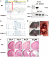
Generation of Dnah5−/− mice. (A) Schematic diagram of the Dnah5 gene locus. The sgRNA sequence is underlined in yellow. The top row shows the wild-type gene sequence. The bottom row shows the gene sequence in the generated Dnah5−/− mice, revealing the deletion of 4 bp enclosed within a red box targeting exon 2 on chromosome 15, leading to a frameshift mutation. (sgRNA = single guide RNA) (B) Results of real-time quantitative PCR. The absence of the Dnah5 gene was confirmed (C) The pattern of the 4 bp deletion in Dnah5−/− mice compared to wild-type mice was demonstrated using pyrosequencing. (D) The head shapes of mice on day five. The top image shows a representative wild-type mouse, while the bottom image shows a representative Dnah5−/− mouse, demonstrating changes in the head shape characteristic of hydrocephalus model mice. In the adjacent image, the Dnah5−/− mouse shows situs inversus, indicating a reversal in the position of internal organs such as the heart, spleen (white arrow), and liver. Each mouse was photographed on 5 mm grid paper. (E) Coronal sections stained with HE from wild-type and Dnah5−/− mice were compared to assess the size of the lateral ventricle anterior horn with age. The size of the ventricles progressively increased day by day. Scale bars = 500 μm.
Figure 2
Figure 2
Ciliary structure and CSF flow. (A) Observation of cilia under electron microscopy in mice on day three. In the TEM (top, scale bar = 20 nm), a horizontal section of the cilia can be observed. The outer dynein arm structures of the peripheral doublet microtubules, indicated by arrows in the illustration, were absent in Dnah5−/− mice, but not in wild-type mice. In the SEM (bottom, scale bar = 5 μm), the ventricular wall observed in Dnah5−/− mice exhibited irregular ciliary extension compared to the wild-type mice. (B) Confirmation of cerebral aqueductal patency in 4-day-old Dnah5−/− mice following injection of the DiI fluorescent dye into the lateral ventricle. The numbered slices 1 to 4 in the illustration represent the locations of coronal brain sections, with slice 1 indicating the anterior horn of the lateral ventricle, slices 2 and 3 depicting the cerebral aqueduct, and slice 4 representing the fourth ventricle. DiI reached the fourth ventricle, indicating patency of the cerebral aqueduct. However, simultaneous observation revealed an already enlarged lateral ventricle. (C) Following immediate extraction of brains from 10-day-old wild-type and Dnah5−/− mice, coronal sections were created, and microbeads were inserted into the ventricles to track their trajectories. In wild-type mice, microbeads exhibited movement with observed vortex flow inside the ventricle. In contrast, no movement of the microbeads was observed in Dnah5−/− mice. The variation in color of the spots in the tracked trajectories represents Z-axis positions, enabling visual evaluation of how the particles move.
Figure 3
Figure 3
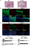
Effects of Dnah5 gene deficiency on cortex. (A) Immunostaining using Neuro Trace (green) and Hoechst 33342 (blue) was performed on the entire six layers of cerebral cortex (100 μm width) within the motor area of the frontal lobe in the brains of 3-day-old wild-type and Dnah5−/− mice (left). Cell counts (n = 8) revealed a reduction of approximately 25% in the number of neurons in the Dnah5−/− mouse cortex compared to the wild-type mouse cortex (right graph, *** p < 0.001). (B) Staining with anti-dynein antibody (red) and Hoechst 33342 (blue) was performed on wild-type and Dnah5−/− mice, capturing images at weak, moderate, and strong magnifications. In wild-type mice, the anti-dynein antibody distinctly stained the cytoplasmic region corresponding to the neuronal axons in the cerebral cortex. Conversely, staining was lower in the Dnah5−/− mouse. Top panels, scale bar = 500 μm; bottom panels, scale bar = 50 μm. (C) Western blotting confirmed a 56.3% reduction in dynein protein expression in the cerebral cortex of Dnah5−/− mice compared to wild-type mice.
Figure 4
Figure 4
Effects on the ependymal and subependymal zone layers. (A) Comparison of the structures of the ventricular wall (ependymal layer and SVZ) between wild-type and Dnah5−/− mice in the brain ventricular wall, as indicated by the white boxes in the figure. Scale bar = 200 μm. (B) Double immunostaining was performed using Neuro Trace and nestin. Neuro Trace staining revealed a decrease in cell density in the ventricular wall of Dnah5−/− mice compared to wild-type mice. Nestin staining also exhibited reduced intensity in Dnah5−/− mice. Scale bar = 20 μm. (C) Double immunostaining was conducted with N-cadherin and nestin. Similarly to nestin staining, N-cadherin staining showed a reduced intensity in Dnah5−/− mice. (Scale bar = 20 μm) (D) Western blotting confirmed a 31.7% decrease in nestin protein expression and a 33.3% reduction in N-cadherin protein expression in Dnah5−/− mice compared to wild-type mice.

References

    1. Kahle K.T., Kulkarni A.V., Limbrick D.D., Jr., Warf B.C. Hydrocephalus in children. Lancet. 2016;387:788–799. doi: 10.1016/S0140-6736(15)60694-8. - DOI - PubMed
    1. Cinalli G., Spennato P., Nastro A., Aliberti F., Trischitta V., Ruggiero C., Mirone G., Cianciulli E. Hydrocephalus in aqueductal stenosis. Child Nerv. Syst. 2011;27:1621–1642. doi: 10.1007/s00381-011-1546-2. - DOI - PubMed
    1. Vinchon M., Rekate H., Kulkarni A.V. Pediatric hydrocephalus outcomes: A review. Fluids Barriers CNS. 2012;9 doi: 10.1186/2045-8118-9-18. - DOI - PMC - PubMed
    1. Varagur K., Sanka S.A., Strahle J.M. Syndromic Hydrocephalus. Neurosurg. Clin. N. Am. 2022;33:67–79. doi: 10.1016/j.nec.2021.09.006. - DOI - PMC - PubMed
    1. Kousi M., Katsanis N. The Genetic Basis of Hydrocephalus. Annu. Rev. Neurosci. 2016;39:409–435. doi: 10.1146/annurev-neuro-070815-014023. - DOI - PubMed

Publication types

MeSH terms

LinkOut - more resources