. 2024 Nov 15;16(22):3836.
doi: 10.3390/cancers16223836.

Novel PP2A-Activating Compounds in Neuroblastoma

Affiliations

Novel PP2A-Activating Compounds in Neuroblastoma

Nazia Nazam et al. Cancers (Basel). .

Abstract

Background: Neuroblastoma (NB) remains one of the deadliest pediatric solid tumors. Recent advancements aimed at improving outcomes have been insufficient, and patients with high-risk NB continue to have a poor prognosis. Protein phosphatase 2A (PP2A) is a tumor suppressor protein downregulated in many cancers, including NB. PP2A activation has been shown to affect the malignant phenotype in other solid tumors. The present studies aim to investigate the effects of two novel PP2A activators as a NB therapeutic.

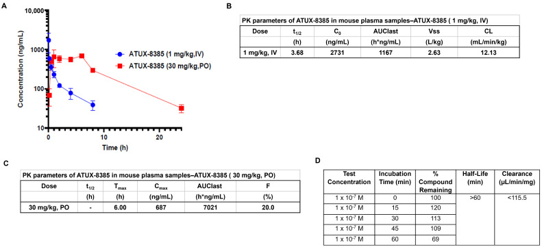
Methods: Four established NB cell lines and a patient-derived xenoline were utilized to study the effect on cell viability, proliferation, motility, and in vivo tumor growth using two novel tricyclic sulfonamide PP2A activators, ATUX-3364 and ATUX-8385.

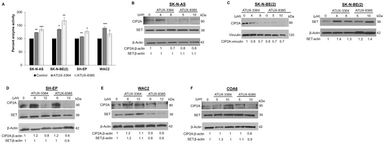
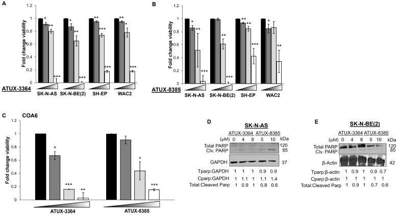
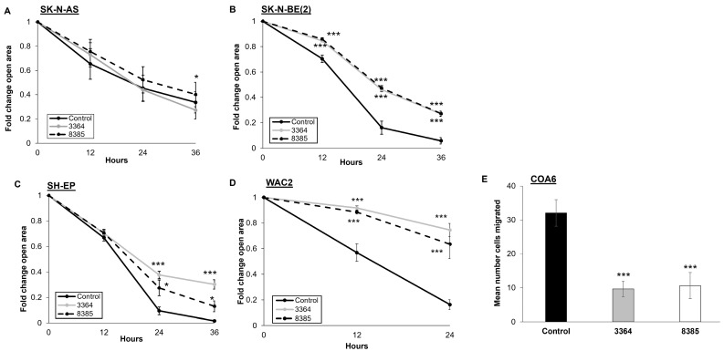
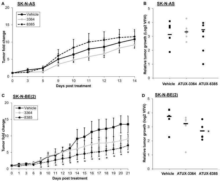
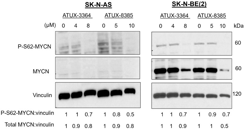
Results: ATUX-3364 and ATUX-8385 increased PP2A activity. These PP2A activators led to decreased viability, proliferation, and motility of NB cells. Treatment of animals bearing NB tumors with ATUX-3364 or ATUX-8385 resulted in decreased tumor growth in MYCN-amplified SK-N-BE(2) tumors. At the molecular level, PP2A-based reactivation led to dephosphorylation of MYCN-S62 and decreased MYCN protein expression.

Conclusions: PP2A activators decreased NB cell viability, proliferation, and motility. In vivo experiments show that PP2A activators have more significant effects on tumorigenesis in MYCN-amplified tumors. Finally, phosphorylation of MYCN protein was decreased following treatment with novel sulfonamide PP2A activators. These data and mechanistic insights may be useful for developing new PP2A-based therapies that target MYCN for the treatment of NB.

Keywords: ATUX-3364; ATUX-8385; neuroblastoma; protein phosphatase 2A.


Conflict of interest statement

M.O. is the inventor of ATUX-3364 and ATUX-8385 and the founder of Atux Iskay, LLC. The other authors declare no competing financial interests in relation to the work described.

Figures

Figure 1
Figure 1
Pharmacokinetic studies for ATUX-8385. (AC) Pharmacokinetic studies were completed by Eurofins. ATUX-8385 was administered at 1 mg/kg intravenously (IV) or 30 mg/kg oral (PO) to mice. The plasma samples were collected, processed using acetonitrile precipitation, and analyzed by LC-MS/MS. (A) Plasma concentrations were sustained longer with PO administration. (B,C) Pharmacokinetic data in tabular form for IV and PO dosing of ATUX-8385. (D) Pharmacokinetic data for in vitro metabolism for ATUX-8385. t1/2 = half-life; C0 = concentration at time zero; AUClast = area under the curve from time of administration up to the time of the last quantifiable concentration; Vss = steady-state volume of distribution; CL = clearance; Tmax = time to maximum concentration; Cmax = maximum concentration; F = bioavailability.
Figure 2
Figure 2
ATUX-3364 and ATUX-8385 affect PP2A enzyme activity. SK-N-AS, SK-N-BE(2), SH-EP, or WAC2 cells were treated with ATUX-3364 or ATUX-8385 for 24 h and PP2A activity measured. Percent PP2A enzyme activity was compared to that of untreated (control) cells. (A) PP2A activation was significantly increased in SK-N-AS with ATUX-3364 or ATUX-8385. Similarly, PP2A was significantly activated in the three other cell lines with both compounds. There was no significant difference in PP2A activation between the two compounds in any of the cell lines (B). In SK-N-AS cells, expression of CIP2A was decreased at higher doses and SET expression was unchanged following ATUX-3364 or ATUX-8385 treatment. (C) Expression of SET (left panel) was increased in SK-N-BE(2) cells and CIP2A decreased (right panel) after treatment. (D) Treatment with ATUX-3364 or ATUX-8385 led to decreased CIP2A expression but did not alter SET expression in SH-EP cells. (E) Treatment of WAC2 cells with ATUX-8385 decreased CIP2A and SET expression but ATUX-3364 increased these proteins at higher doses. (F) CIP2A expression was increased with the highest dose of ATUX-8385 but SET expression was variably affected in the NB PDX COA6 cells. Data reported as mean fold change ± standard error of the mean (SEM), and experiments were repeated with at least three biologic replicates. * p ≤ 0.05, ** p ≤ 0.01, *** p ≤ 0.001. The uncropped bolts are shown in Supplementary Materials.
Figure 3
Figure 3
ATUX-3364 and ATUX-8385 decreased NB viability. Cells from established NB cell lines SK-N-AS, SK-N-BE(2), SH-EP, and WAC2 were treated with increasing doses of (A) ATUX-3364 (0–20 µM) or (B) ATUX-8385 (0–20 µM) for 24 h. Both compounds significantly decreased viability in all cell lines. (C) COA6 human NB PDX cells were treated with increasing doses of ATUX-3364 or ATUX-8385 (0–25 µM) for 24 h. Both compounds decreased viability. Immunoblotting was performed on whole cell lysates from (D) SK-N-AS and (E) SK-N-BE(2) NB cells following treatment with ATUX-3364 or ATUX-8385. Cleaved PARP (85 kDa) was increased, and total PARP (120 kDa) decreased in SK-N-AS and SK-N-BE(2) whole cell lysates. Data are reported as mean fold change ± SEM, and experiments were repeated with at least three biological replicates. * p ≤ 0.05, ** p ≤ 0.01, *** p ≤ 0.001. The uncropped bolts are shown in Supplementary Materials.
Figure 4
Figure 4
ATUX-3364 and ATUX-8385 decreased NB motility. Migration was examined using monolayer wound healing (scratch) assay for established NB cell lines. After a sterile scratch, plates were treated with ATUX-3364 or ATUX-8385, and micrographs of the plates were obtained. For quantification of open wound area, ImageJ MRI Wound Healing Tool (https://imagej.net, accessed on 26 November 2021) was used. (A) Migration of SK-N-AS cells was only affected by ATUX-8385 (4 μM). (B) Treatment of SK-N-BE(2) cells with ATUX-3364 or ATUX-8385 (4 μM) significantly decreased migration. (C) SH-EP and (D) WAC2 cells were treated with ATUX-3364 or ATUX-8385 (12 μM). Migration in both cell lines was significantly decreased following treatment. (E) Migration was assessed in COA6 PDX cells using modified Boyden chamber assays. Treatment with ATUX-3364 and ATUX-8385 (6 μM) significantly decreased COA6 migration. Data are reported as mean fold change in area of open area or mean number of cells migrated ± SEM, and experiments were repeated with at least three biological replicates. * p ≤ 0.05, *** p ≤ 0.001.
Figure 5
Figure 5
ATUX-8385 decreased SK-N-BE(2) tumor growth in vivo. SK-N-AS or SK-N-BE(2) cells were injected into the right flank of female athymic nude mice. When tumor volume reached 100 mm3, animals were randomized into groups of three to receive either vehicle, ATUX-3364, or ATUX-8385 (50 mg/kg). (A,B) Animals bearing SK-N-AS tumors (n = 7 in vehicle, n = 6 in ATUX-3364, n = 6 in ATUX-8385 treatment groups) were treated for 14 days. There was no significant difference in change in tumor volume or relative tumor growth with either compound. (C,D) Animals bearing SK-N-BE(2) tumors (n = 5 in vehicle, n = 6 in ATUX-3364, and n = 7 in ATUX-8385 treatment groups) were treated for 21 days. There was a significant decrease in tumor volume and relative tumor growth with ATUX-8385 treatment. Data are reported as mean fold change in tumor volume ± SEM. * p ≤ 0.05.
Figure 6
Figure 6
ATUX-3364 and ATUX-8385 decreased serine phosphorylation of MYCN. SK-N-AS (MYCN-non-amplified) and SK-N-BE(2) (MYCN-amplified) cells were treated with increasing concentrations of ATUX-3364 or ATUX-8385. Whole cell lysates were prepared, and immunoblotting completed for phosphorylated MYCN (P-S62) and total MYCN protein expression. Vinculin was assayed as a loading control. Serine 62 phosphorylation of MYCN was decreased with treatment with both drugs. Total MYCN protein expression was also decreased following treatment. The uncropped bolts are shown in Supplementary Materials.

References

    1. Cohn S.L., Pearson A.D.J., London W.B., Monclair T., Ambros P.F., Brodeur G.M., Faldum A., Hero B., Iehara T., Machin D., et al. The International Neuroblastoma Risk Group (INRG) Classification System: An INRG Task Force Report. J. Clin. Oncol. 2009;27:289–297. doi: 10.1200/JCO.2008.16.6785. - DOI - PMC - PubMed
    1. Smith V., Foster J. High-Risk Neuroblastoma Treatment Review. Children. 2018;5:114. doi: 10.3390/children5090114. - DOI - PMC - PubMed
    1. Colon N.C., Chung D.H. Neuroblastoma. Adv. Pediatr. 2011;58:297–311. doi: 10.1016/j.yapd.2011.03.011. - DOI - PMC - PubMed
    1. Matthay K.K., Maris J.M., Schleiermacher G., Nakagawara A., Mackall C.L., Diller L., Weiss W.A. Neuroblastoma. Nat. Rev. Dis. Primers. 2016;2:16078. doi: 10.1038/nrdp.2016.78. - DOI - PubMed
    1. Weiss W.A. Targeted Expression of MYCN Causes Neuroblastoma in Transgenic Mice. EMBO J. 1997;16:2985–2995. doi: 10.1093/emboj/16.11.2985. - DOI - PMC - PubMed

LinkOut - more resources