. 2024 Nov 13;11(11):1377.
doi: 10.3390/children11111377.

Pediatric Cranial Vault Lesions: A Tailored Approach According to Bony Involvement

Affiliations

Pediatric Cranial Vault Lesions: A Tailored Approach According to Bony Involvement

Arianna Barbotti et al. Children (Basel). .

Abstract

Background: Cranial vault lesions are common in children, with dermoid and epidermoid cysts being the most frequent. Management is debated due to their slow growth, but early resection can prevent complications and provide a definitive histological diagnosis, which is sometimes linked to systemic diseases.

Methods: A retrospective study of children treated surgically for cranial vault tumors from January 2011 to April 2023 was conducted. The data collected included age, gender, symptoms, comorbidities, lesion location, radiological features, surgical techniques, histopathology, and recurrence rates.

Results: Eighty-eight children (mean age: 3.5 years, mean tumor size: 1.21 cm) underwent surgery. The most common locations were the frontal and occipital bones. The main diagnoses were dermoid cysts, myofibroma, and Langerhans cell histiocytosis. Gross total resection was achieved in 64 cases with simple skin incisions. In 13 cases, small cranioplasties with bone cement were used. Craniotomy and cranioplasty with autologous bone grafting were performed for 11 patients with lesions larger than 2 cm and full skull thickness erosion.

Conclusions: Early resection is recommended for complete removal with minimally invasive surgery and to ensure histological diagnosis. For lesions larger than 2 cm with full skull erosion, cranioplasty with autologous bone is the preferred technique.

Keywords: Langerhans cell histiocytosis; cranial vault; cranioplasty; dermoid.


Conflict of interest statement

The authors declare no conflicts of interest.

Figures

Figure 1
Figure 1
Surgical decision workflow according to lesion dimension and bone involvement.
Figure 2
Figure 2
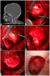
Complete removal of a frontal dermoid. In (A), the sagittal CT scans show a through-and-through bony defect slightly anterior to the bregmatic fontanel. After the skin incision, a subperiosteal lump was identified (B), represented by a well-encapsulated red–yellow mass eroding the whole thickness of the skull (C). The lesion, which was compatible with a dermoid, was enucleated (D). (E) shows an intraoperative view of frontal bone erosion after the en bloc removal of the dermoid cyst (F).
Figure 3
Figure 3
MRI and CT scans showing a parietal bone lesion extending in the epidural space (A,B); intraoperative appearance (C); bone-splitting procedure with piezo saw (D); final reconstruction (E); postoperative CT scan showing the closure of the bone defect (F).
Figure 4
Figure 4
How to manage complications in wider tumors: a multistep approach. The three-dimensional CT scan depicts (A) a right frontotemporal intraosseous lesion, with intracranial extension ((B)—coronal CT scan). Surgery was performed, based only on tumor removal via craniectomy, without cranioplasty, to obtain re-ossification during growth; the histopathological diagnosis was of melanocytic neuroectodermal tumor. However, due to the onset of a large fluctuant lump (C), a second surgery with allograft bone was achieved (D): the CT shows the bone flap fixed with absorbable plates. After three years, central resorption of the bone graft was revealed (E), with progressive enlargement of the bone defect (F). Considering the currently sufficient thickness of the skull vault bone, the residual allograft was removed, and autologous bone grafting obtained with the “splitting” technique, secured with nonabsorbable plates, was performed (G). The last CT examination (H), 12 years after the first surgery, confirmed the absence of tumor recurrence and the complete re-ossification of the bone defect.

References

    1. Ruge J.R., Tomita T., Naidich T.P., Hahn Y.S., McLone D.G. Scalp and Calvarial Masses of Infants and Children. Neurosurgery. 1988;22:1037–1042. doi: 10.1227/00006123-198806010-00011. - DOI - PubMed
    1. Barnett R.R., Piazza M.G., Elton S.W. Pediatric Neurosurgery in Primary Care: Masses of the Scalp and Skull in Children. Pediatr. Clin. N. Am. 2021;68:743–757. doi: 10.1016/j.pcl.2021.04.003. - DOI - PubMed
    1. Cummings T.J., George T.M., Fuchs H.E., McLendon R.E. The Pathology of Extracranial Scalp and Skull Masses in Young Children. Clin. Neuropathol. 2004;23:33–43. - PubMed
    1. Sorenson E.P., Powel J.E., Rozzelle C.J., Tubbs R.S., Loukas M. Scalp Dermoids: A Review of Their Anatomy, Diagnosis, and Treatment. Child’s Nerv. Syst. 2013;29:375–380. doi: 10.1007/s00381-012-1946-y. - DOI - PubMed
    1. Skadorwa T., Ciszek B. Clinical Characteristics of Benign Pediatric Cranial Vault Tumors: Surgical Considerations Based on 100 Cases. Pediatr. Neurosurg. 2016;52:13–19. doi: 10.1159/000448045. - DOI - PubMed