. 2024 Oct 24;12(11):2439.
doi: 10.3390/biomedicines12112439.

Neutrophils and Lymphocytes: Yin and Yang of Lung Fibrosis and Patient Outcome in Diffuse Interstitial Lung Diseases

Affiliations

Neutrophils and Lymphocytes: Yin and Yang of Lung Fibrosis and Patient Outcome in Diffuse Interstitial Lung Diseases

Erika M Novoa-Bolivar et al. Biomedicines. .

Abstract

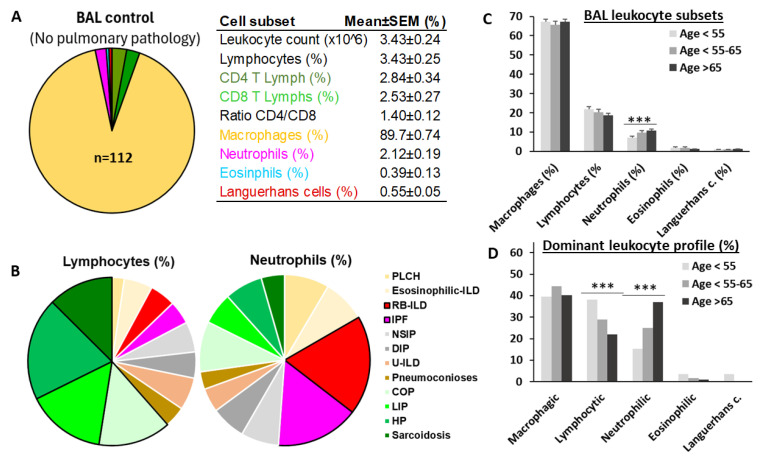
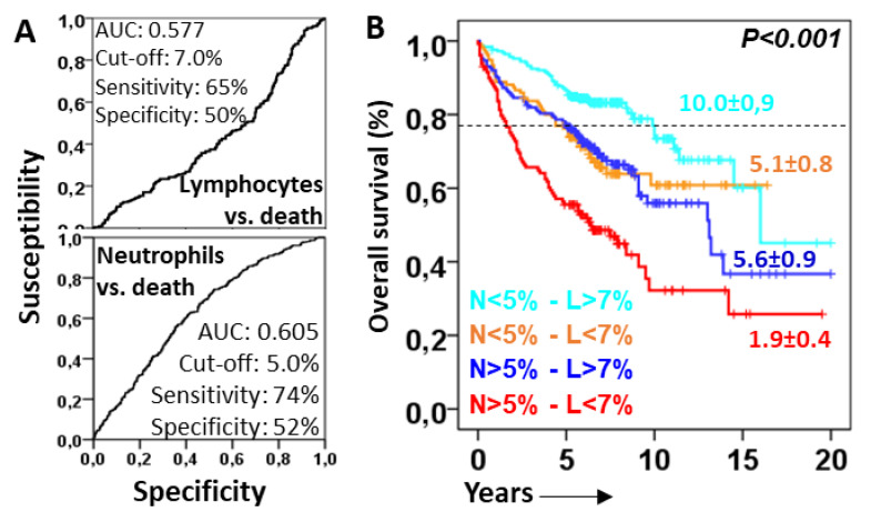
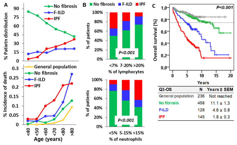
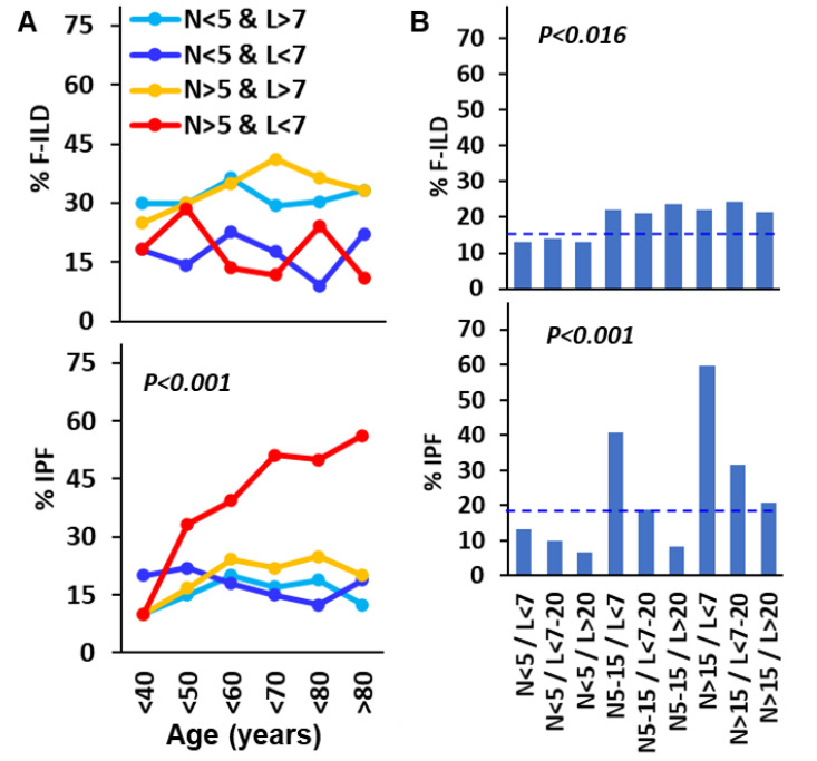
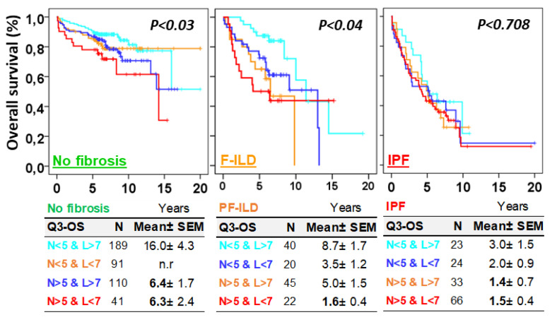
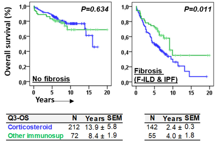
Objective: Antifibrotics can improve the outcome of patients with idiopathic pulmonary fibrosis (IPF) and other fibrosing interstitial lung diseases (F-ILDs), but predictive biomarkers at diagnosis are needed to guide the use of immunomodulating and antifibrotic therapies. Methods: Flow cytometry quantification of lymphocytes and neutrophils in bronchoalveolar lavage (BAL) of 145 IPFs, 561 non-IPF-ILDs (125 F-ILDs), and 112 BAL controls were retrospectively correlated with the incidence of fibrosis and third-quartile overall survival (Q3-OS). Results: The incidence of IPF was directly proportional (9.6%, 22.2%, and 42.6%, p < 0.001) to BAL neutrophil counts (<5%, 5-15%, and >15%), but inversely proportional (34.1%, 18.6%, and 8.8%, p < 0.001) to BAL lymphocyte counts (<7%, 7-20%, and >20%). Elevated neutrophils (>5%) with low lymphocytes (<7%) were associated with an increasingly higher incidence of IPF (10.0-56.3%, p < 0.001) in patients aged 40 to 80, compared to the rest of patients (13.0-17.1%). Lymphocytes >20% compared to lymphocytes <7% strongly protected patients with neutrophils >15% (59.7% vs. 20.7%, p < 0.001) from IPF. In contrast, the incidence of F-ILD was not clearly related to BAL lymphocyte/neutrophil counts. Although, IPF and F-ILD showed a shorter Q3-OS (1.8 ± 0.3 and 4.6 ± 0.8 years; p < 0.001) than non-fibrotic-ILDs (11.1 ± 1.3 years), lymphocyte and neutrophil counts were associated with a longer and shorter Q3-OS of non-fibrotic-ILDs (p < 0.03) and F-ILDs (p < 0.04), respectively, but not with a Q3-OS of IPF patients (p < 0.708). Corticosteroids in patients with fibrosis showed a shorter Q3-OS than other immunomodulators (2.4 ± 0.3 vs. 4.0 ± 1.8 years, p = 0.011). Conclusions: Accurate counting of BAL lymphocytes and neutrophils by flow cytometry in ILD patients at diagnosis could help guide immunomodulatory and antifibrotic therapies.

Keywords: BAL lymphocyte and neutrophils; flow cytometry; idiopathic pulmonary fibrosis; interstitial lung disease; lung fibrosis; patient outcome.


Conflict of interest statement

The authors declare no conflicts of interest.

Figures

Figure 1
Figure 1
Flow cytometry analysis of the leukocyte subsets contained in BAL samples. Cell subsets were selected and colored as follows: lymphocytes in light brown (CD45++ SSClow), macrophages in cyan (DR+ SSHigh), neutrophils in light green (CD66+ CD16+ DR−), eosinophils in magenta (CD16− CD66dim), Langerhans cells in red (CD1a+), T lymphocytes in dark green (CD3+), and B lymphocytes in dark blue (CD19+ CD20+). Additionally, the CD3+CD4+ and CD3+CD8+ T subpopulations were detected. Likewise, the beads contained in the TrueCount tube were identified with a pink color, which was used to calculate the absolute cell count per microliter of the sample. Analysis gates were combined hierarchically and logically as shown on the right side and described at the bottom of the figure.
Figure 2
Figure 2
Lymphocytic ILDs decrease and neutrophilic ILDs increase with aging. (A) Mean values of leukocyte subset in bronchoalveolar lavage (BAL) of individuals without pulmonary pathology; (B) mean BAL lymphocyte and neutrophil values in patients with different types of interstitial lung disease (ILD); (C) mean values of macrophages, lymphocytes, neutrophils, eosinophils, and CD1a+ Langerhans cells in the BAL of total ILD patients according to age, at <55, 55–65, and >65 years; and (D) proportion of patients with lymphocytic (lymphocytes > 20%), neutrophilic (neutrophils > 10%), eosinophilic (eosinophils > 3%), and Langerhans cells (>1.5%) profiles, for patients aged <55, 55–65, and >65. ***, indicate p < 0.001 in the ANOVA test.
Figure 3
Figure 3
Lymphocytes protect from the deleterious effect of lung-infiltrating neutrophils. (A) Receiver operating characteristic curve (ROC) of lymphocytes and neutrophils related to overall survival (OS). Area under the curve (AUC) cut-offs with the highest sensitivity and specificity are shown; (B) Kaplan–Meier and Log-Rank tests for the OS of ILD patients according to the combinations of lymphocytes (above or below 7%) and neutrophils (above or below 5%). The 75th percentile OS is shown for each case.
Figure 4
Figure 4
Incidence of fibrotic ILD (F-ILD), idiopathic pulmonary fibrosis (IPF), and death according to the age and the counts of lymphocytes and neutrophils in the BAL of patients. (A) Incidence of F-ILD, IPF, and death (estimated as the percentage of the sum of deaths divided by the sum of follow-up times of patients) according to the age of ILD patients; (B) incidence of F-ILD and IPF in patients according to the counts of lymphocytes (<7%, 7–20%, or >20%) and neutrophils (<5%, 5–15%, or >15%) in the BAL at diagnosis. p < 0.001 in the Chi-square test; and (C) Kaplan–Meier and Log-Rank tests for overall survival (OS) of the general population, ILD patients without fibrosis, and F-ILD and IPF patients. Pulmonary fibrosis was computed with the presence of reticular changes, traction bronchiectasis, and honeycombing in the radiological study. The 75th percentile OS (Q3–OS) is shown for each case.
Figure 5
Figure 5
Lymphocytes counteract neutrophil-promoted idiopathic pulmonary fibrosis (IPF) during aging. (A) Incidence of F-ILD and IPF according to the age of ILD patients and the combined counts of neutrophils (>5%) and lymphocytes (>7%) in the BAL; (B) incidence of F-ILD and IPF in ILD patients according to the combined counts of neutrophils (<5%, 5–15%, or >15%) and lymphocytes (<7%, 7–20%, or >20%) in the BAL of ILD patients at diagnosis. Pulmonary fibrosis was computed with the presence of reticular changes, traction bronchiectasis, and honeycombing in the radiological study p estimated using the Chi-square test. Dashed lines indicate the mean incidence of fibrosis in total patients.
Figure 6
Figure 6
Neutrophil and lymphocyte counts were associated with the overall survival (OS) of ILD patients without fibrosis and with pulmonary fibrosis associated with ILDs (F-ILDs), but not with the OS of patients with idiopathic pulmonary fibrosis (IPF). Kaplan–Meier and Log-Rank tests for the OS of ILD patients according to the combinations of lymphocytes (above or below 7%) and neutrophils (above or below 5%) and the type of fibrotic process (F-ILD or IPF). Pulmonary fibrosis was computed with the presence of reticular changes, traction bronchiectasis, and honeycombing in the radiological study.
Figure 7
Figure 7
Immunomodulating therapies were differentially associated with overall survival in patients with or without fibrosis. Kaplan–Meier and Log-Rank tests for the overall survival (OS) of ILD patients according to the type of immunomodulatory treatment, corticosteroids vs. other immunosuppressants (rituximab, azathioprine, mycophenolate mofetil, cyclophosphamide, or tacrolimus), and the presence of fibrosis. The 75th percentile of the OS (Q3–OS) is shown for each case.

References

    1. Bagnato G., Harari S. Cellular interactions in the pathogenesis of interstitial lung diseases. Eur. Respir. Rev. 2015;24:102–114. doi: 10.1183/09059180.00003214. - DOI - PMC - PubMed
    1. Moss B.J., Ryter S.W., Rosas I.O. Pathogenic Mechanisms Underlying Idiopathic Pulmonary Fibrosis. Annu. Rev. Pathol. Mech. Dis. 2022;17:515–546. doi: 10.1146/annurev-pathol-042320-030240. - DOI - PubMed
    1. Lederer D.J., Martinez F.J. Idiopathic Pulmonary Fibrosis. N. Engl. J. Med. 2018;378:1811–1823. doi: 10.1056/NEJMra1705751. - DOI - PubMed
    1. Cottin V., Hirani N.A., Hotchkin D.L., Nambiar A.M., Ogura T., Otaola M., Skowasch D., Park J.S., Poonyagariyagorn H.K., Wuyts W., et al. Presentation, diagnosis and clinical course of the spectrum of progressive-fibrosing interstitial lung diseases. Eur. Respir. Rev. 2018;27:180076. doi: 10.1183/16000617.0076-2018. - DOI - PMC - PubMed
    1. Wells A.U., Flaherty K.R., Brown K.K., Inoue Y., Devaraj A., Richeldi L., Moua T., Crestani B., Wuyts W.A., Stowasser S., et al. Nintedanib in patients with progressive fibrosing interstitial lung diseases—Subgroup analyses by interstitial lung disease diagnosis in the INBUILD trial: A randomised, double-blind, placebo-controlled, parallel-group trial. Lancet Respir. Med. 2020;8:453–460. doi: 10.1016/S2213-2600(20)30036-9. - DOI - PubMed

LinkOut - more resources