. 2024 Nov 6;14(11):1412.
doi: 10.3390/biom14111412.

Restored Collagen VI Microfilaments Network in the Extracellular Matrix of CRISPR-Edited Ullrich Congenital Muscular Dystrophy Fibroblasts

Affiliations

Restored Collagen VI Microfilaments Network in the Extracellular Matrix of CRISPR-Edited Ullrich Congenital Muscular Dystrophy Fibroblasts

Daniela Benati et al. Biomolecules. .

Abstract

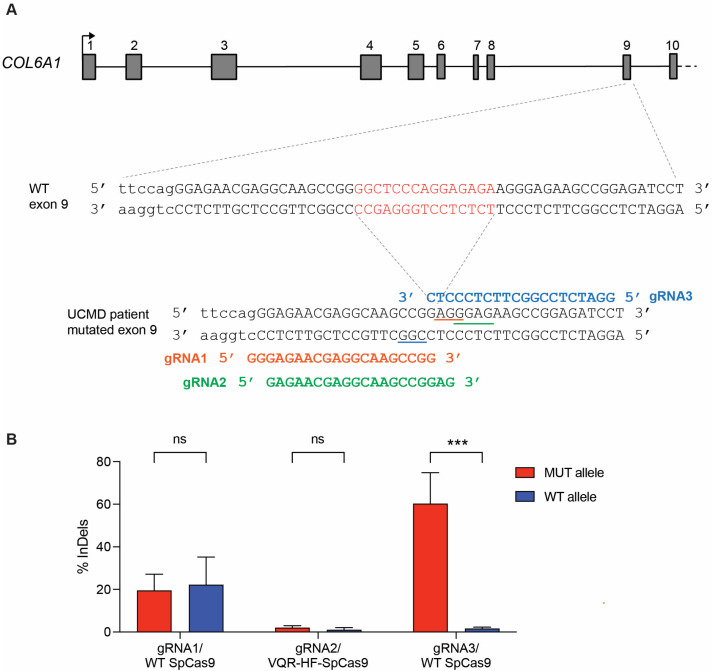
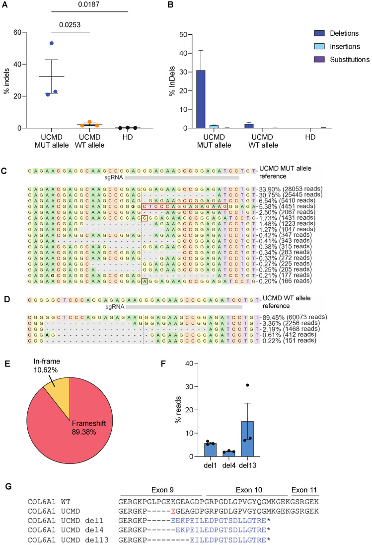
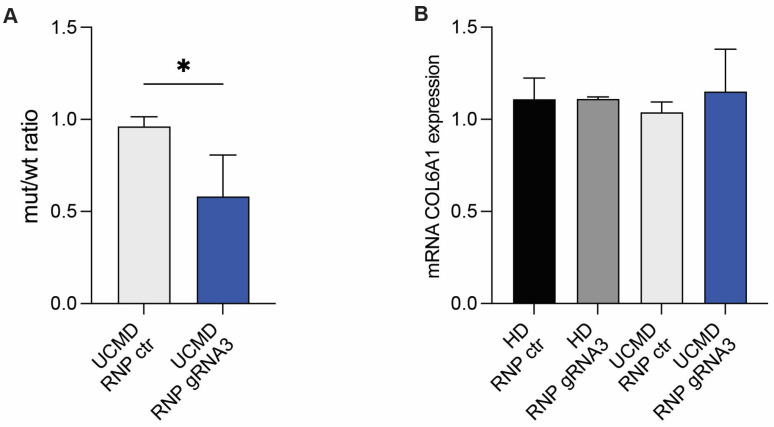
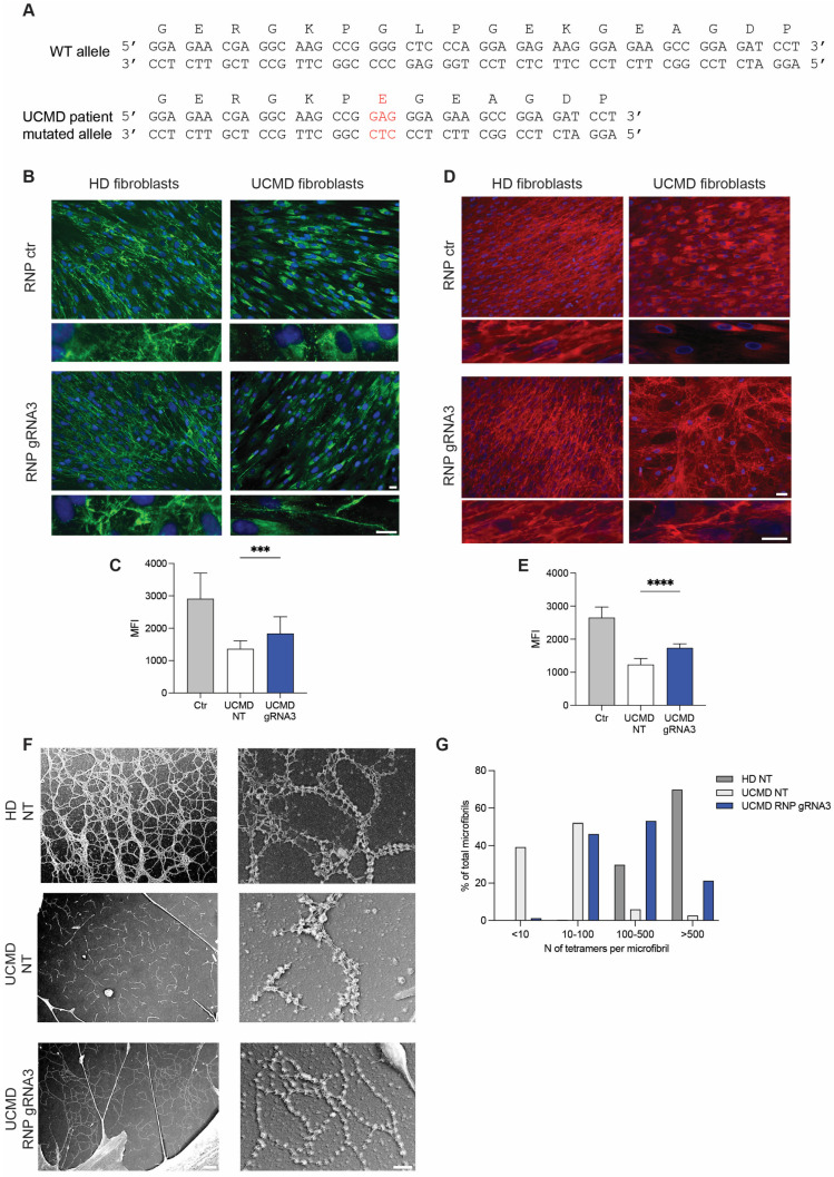
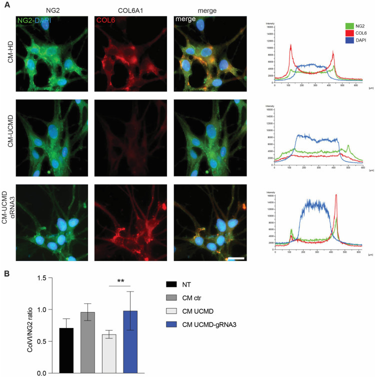
Collagen VI is an essential component of the extracellular matrix (ECM) composed by α1, α2 and α3 chains and encoded by COL6A1, COL6A2 and COL6A3 genes. Dominant negative pathogenic variants in COL6A genes result in defects in collagen VI protein and are implicated in the pathogenesis of muscular diseases, including Ullrich congenital muscular dystrophy (UCMD). Here, we designed a CRISPR genome editing strategy to tackle a dominant heterozygous deletion c.824_838del in exon 9 of the COL6A1 gene, causing a lack of secreted collagen VI in a patient's dermal fibroblasts. The evaluation of efficiency and specificity of gene editing in treating patient's fibroblasts revealed the 32% efficiency of editing the mutated allele but negligible editing of the wild-type allele. CRISPR-treated UCMD skin fibroblasts rescued the secretion of collagen VI in the ECM, which restored the ultrastructure of the collagen VI microfibril network. By using normal melanocytes as surrogates of muscle cells, we found that collagen VI secreted by the corrected patient's skin fibroblasts recovered the anchorage to the cell surface, pointing to a functional improvement of the protein properties. These results support the application of the CRISPR editing approach to knock out COL6A1 mutated alleles and rescue the UCMD phenotype in patient-derived fibroblasts.

Keywords: CRISPR/Cas9; allele-specific gene editing; collagen VI; collagen VI-related disorders; patient-derived fibroblasts.


Conflict of interest statement

The authors declare no conflicts of interests.

Figures

Figure 1
Figure 1
CRISPR/Cas9 allele-specific targeting of c.824_838del COL6A1 dominant variant. (A) Schematic representation (not in scale) of the first 10 translated exons of COL6A1 gene Each exon is numbered from 1 to 10. The arrow above the first exon indicates the transcription start site. The sequence of the genomic region surrounding exon 9 in the wild-type (WT) allele and UCMD allele carrying the heterozygous c.824_838del variant are shown (exon 9 in capital letters, the last nucleotides of intron 8 in lower case). The 15-nucleotide deletion is highlighted in red on the WT sequence. The picture illustrates gRNA1 (red), gRNA2 (green) and gRNA3 (light blue) targeting the mutation. PAM sequences are underlined. (B) Frequency of indels determined by TIDE analysis on HEK 293T cells transfected with effector plasmids for SpCas9 variants and gRNA and plasmids carrying the WT or UCMD allele sequences. The experiment was performed in triplicate and is presented as mean ± SEM. *** p < 0.001; ns, not significant.
Figure 2
Figure 2
NGS data analysis of UCMD patient cells electroporated with RNP/gRNA3. (A) Frequency of indels in exon 9 of WT or mutant allele (MUT) from UCMD fibroblasts or from HD fibroblasts. The experiment was performed in triplicate and is presented as mean ± SEM. * p < 0.05. (B) Type of indels, and their relative percentage, generated in WT or mutant allele (MUT) from UCMD fibroblasts or from HD fibroblasts. (C,D) CRISPResso2 graphic representation of the distribution of identified alleles around the cleavage site of the gRNA3 on UCMD mutated allele or on WT allele. The top sequence is the unmodified reference. Substitutions are shown in bold font. Red rectangles highlight inserted sequences. Horizontal dashed lines indicate deleted sequences. The vertical dashed line indicates the predicted cleavage site. A representative experiment is shown. (E) CRISPResso2 analysis of indels generated on UCMD MUT allele leading to frameshift or in-frame mutations. The mean of triplicates is presented in pie chart. (F) Percentages of the three most frequent mutants (del1, del4, del13) identified by CRISPResso2 upon editing the MUT allele of UCMD fibroblasts. (G) Amino acid sequence of exon 9-11 in COL6A1 WT, UCMD c.824_838del variant diagnosed in patient cells and UCMD variants (del1, del4 and del13) identified by CRISPResso2 upon editing the MUT allele of UCMD fibroblasts. The amino acid substitution resulted from the 15-nt deletion in UCMD MUT allele is highlighted in red, while different amino acid sequences resulting from CRISPR-induced frameshift are in blue. Dash indicates deleted codons, and star indicates premature stop codon downstream the canonical one.
Figure 3
Figure 3
Efficient knockdown of mutant allele expression. (A) Measurement of the WT and mutant COL6A1 transcripts by allele-specific ddPCR in UCMD fibroblasts treated with RNP-gRNA3 or control RNP (RNP ctr). Mutant/WT COL6A1 allele ratio in UCMD patient fibroblasts treated with RNPs is reported in the column chart. Mean ± SD is shown. *, p < 0.05. (B) Measurement of mRNA levels of a total COL6A1 expression by ddPCR in HD and UCMD fibroblasts treated with control RNP (RNP ctr) or RNP-gRNA3. The results of four experiments are shown as mean ± SD.
Figure 4
Figure 4
Secretion of collagen VI in fibroblasts from healthy donor (HD) and UCMD patient after treatment with RNP. (A) Nucleotide and amino acid sequence of exon 9 in UCMD patient carrying c.824_838del variant. In red, the insertion of a Glu residue is shown. (B) Immunofluorescence analysis with antibody specific for collagen VI (MAB1944) of fibroblasts from HD or UCMD patient, treated with control RNP (RNP ctr) or RNP-gRNA3. The experiment was performed in quadruplicate. A representative experiment is shown. Enlarged image insets show the structure of collagen VI microfibrils. Scale bars indicate 20 µm magnification. (C) Mean intensity of collagen VI quantified in fibroblasts treated with RNP-gRNA3. Data represent mean ± SD from analysis of 10 individual field images acquired at 100× original magnification under fluorescence microscopy. *** p < 0.001). (D) Immunofluorescence analysis with collagen VI-specific antibody (polyclonal Fitzgerald) of primary fibroblasts from HD or UCMD patient treated with control RNP (RNP ctr) or RNP-gRNA3. The polyclonal Fitzgerald anti-collagen VI antibody was used to label α1, α2 and α3 chains of collagen VI. A representative experiment is shown. Enlarged image insets show the structure of collagen VI microfibrils. Scale bars indicate 20 µm magnification. (E) Mean intensity of collagen VI quantified in fibroblasts treated with RNP-gRNA3. Data represent mean ± SD from analysis of 10 individual field images acquired at 100× original magnification under fluorescence microscopy. **** p < 0.0001). (F) Ultrastructural analysis by rotary-shadowing electron microscopy of collagen VI microfibrillar network in untreated (NT) HD fibroblasts, UCMD fibroblasts treated with RNP-gRNA3 or not (UCMD NT). (G) Percentage of microfibrils composed by increasing amounts of tetramers in UCMD fibroblasts treated with RNP-gRNA3 or not (UCMD NT) and in control fibroblasts (HD NT).
Figure 5
Figure 5
Restoration of binding of NG2 by collagen VI secreted by UCMD patient after treatment with RNP. (A) Immunofluorescence analysis of NG2 and collagen VI in melanocytes from healthy donor treated with collagen VI-containing conditioned medium (CM) of fibroblasts from HD or UCMD patient untreated or treated with RNP-gRNA3. The experiment was performed in triplicate. Representative mean fluorescence intensity profiles of each sample are shown on the right of each row. A representative experiment is shown. Scale bar indicates 20 µm magnification. (B) COL6/NG2 ratio in melanocytes untreated (NT) or cultivated with conditioned medium (CM) of fibroblasts from HD (ctr), UCMD or UCMD treated with RNP-gRNA3. The experiment was performed in triplicate, and the mean ± SEM is shown. ** p < 0.01.

References

    1. Bernardi P., Bonaldo P. Dysfunction of mitochondria and sarcoplasmic reticulum in the pathogenesis of collagen VI muscular dystrophies. Ann. N. Y. Acad. Sci. 2008;1147:303–311. doi: 10.1196/annals.1427.009. - DOI - PubMed
    1. Cescon M., Gattazzo F., Chen P., Bonaldo P. Collagen VI at a glance. J. Cell Sci. 2015;128:3525–3531. doi: 10.1242/jcs.169748. - DOI - PubMed
    1. Lamande S.R., Bateman J.F. Collagen VI disorders: Insights on form and function in the extracellular matrix and beyond. Matrix Biol. 2018;71–72:348–367. doi: 10.1016/j.matbio.2017.12.008. - DOI - PubMed
    1. Speer M.C., Tandan R., Rao P.N., Fries T., Stajich J.M., Bolhuis P.A., Jobsis G.J., Vance J.M., Viles K.D., Sheffield K., et al. Evidence for locus heterogeneity in the Bethlem myopathy and linkage to 2q37. Hum. Mol. Genet. 1996;5:1043–1046. doi: 10.1093/hmg/5.7.1043. - DOI - PubMed
    1. Camacho Vanegas O., Bertini E., Zhang R.Z., Petrini S., Minosse C., Sabatelli P., Giusti B., Chu M.L., Pepe G. Ullrich scleroatonic muscular dystrophy is caused by recessive mutations in collagen type VI. Proc. Natl. Acad. Sci. USA. 2001;98:7516–7521. doi: 10.1073/pnas.121027598. - DOI - PMC - PubMed

Supplementary concepts

LinkOut - more resources