. 2024 Nov 6;25(22):11943.
doi: 10.3390/ijms252211943.

Direct Effects of the Janus Kinase Inhibitor Baricitinib on Sensory Neurons

Affiliations

Direct Effects of the Janus Kinase Inhibitor Baricitinib on Sensory Neurons

Enrique Vazquez et al. Int J Mol Sci. .

Abstract

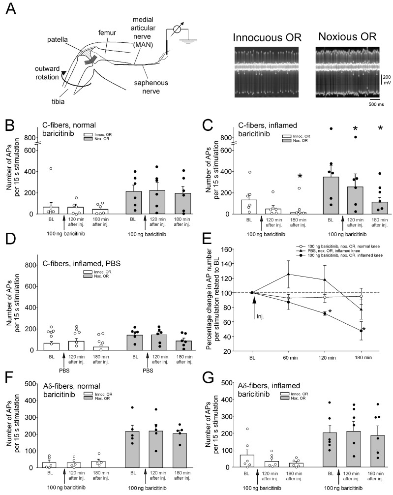
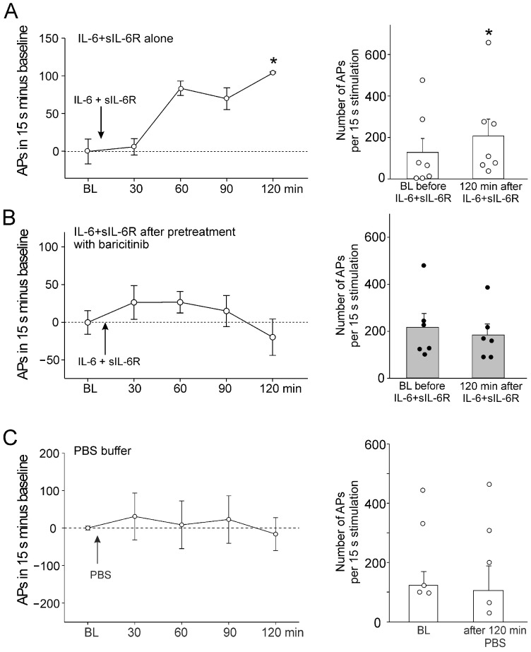
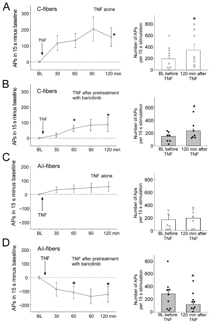
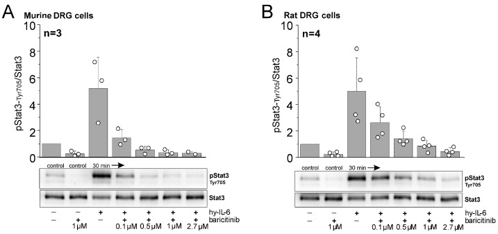
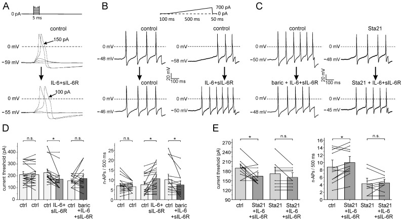
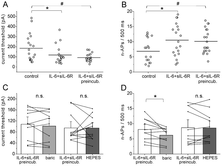
Therapeutically, the Janus kinase (Jak) 1/Jak2 inhibitor baricitinib reduces the pathology of rheumatoid arthritis and may also reduce pain. Here, we investigated whether baricitinib directly affects joint nociceptors. We recorded action potentials from nociceptive C- and A∂-fibers of the normal and inflamed knee joint in anesthetized rats to monitor their responses to innocuous and noxious joint rotation. In isolated and cultured dorsal root ganglion (DRG) neurons, we examined Stat3 activation using Western blots and monitored excitability using patch-clamp recordings. Intra-articular injection of baricitinib did not alter C- and A∂-fiber responses to innocuous and noxious rotations of the normal knee but reduced C-fiber responses to these stimuli in inflamed joints. Baricitinib prevented the increase in C-fiber responses to joint rotation evoked by interleukin (IL)-6 plus soluble interleukin-6 receptor (sIL-6R) but not the increase evoked by TNF. In DRG neurons, baricitinib blocked Stat3 activation by hyper-IL-6, and baricitinib or the Stat3 inhibitor Sta21 prevented induction of hyperexcitability by IL-6 plus sIL-6R. Thus, neuronal Jaks are involved in the generation of C-fiber hyperexcitability induced by inflammation and IL-6. Pain reduction by baricitinib may result, at least in part, from direct effects on joint nociceptors.

Keywords: DRG neurons; Jak inhibitor; Stat3; TNF; baricitinib; interleukin-6; joint pain; neuronal Jak; nociceptor; pain sensitization.


Conflict of interest statement

The authors declare no conflicts of interest. The funders had no role in the design of the study, in the collection, analyses, or interpretation of data, in the writing of the manuscript, or in the decision to publish the results.

Figures

Figure 1
Figure 1
Effect of intra-articular injection of baricitinib on joint nociceptor responses to innocuous and noxious outward rotation (OR) of the normal and inflamed knee joint. (A) Experimental setup for in vivo recordings from joint nociceptors in anesthetized rats (left) and typical action potentials of a single nerve fiber evoked by innocuous and noxious OR (right). (B) No change in response magnitude of normal knee joint C-fibers (n = 6) to innocuous and noxious OR by baricitinib application (arrows). Fibers were continuously recorded for 180 min. (C) Reduction of C-fiber responses (n = 6) of the acutely inflamed joint to innocuous and noxious OR by baricitinib application. (D) No significant change of C-fiber responses (n = 6) to innocuous and noxious OR of the acutely inflamed joint by PBS. (E) Summary of the effects shown in (BD). Baseline values before baricitinib or PBS injection were normalized to 100%. (F) No change in A∂-fiber responses (n = 5) of the normal knee joint to innocuous and noxious OR by baricitinib. (G) No change in A∂-fiber responses (n = 6) of the inflamed knee joint to innocuous and noxious OR by baricitinib. White circles and black bullets indicate single data points from individual fibers. * p < 0.05, Wilcoxon matched-pairs signed-rank test.
Figure 2
Figure 2
Effect of baricitinib on induction of C-fiber hyperexcitability by IL-6 + sIL-6R. (A) Persistent increase in C-fiber responses (n = 7) to noxious OR by intra-articular injection of IL-6 + sIL-6R (arrow). The right columns show responses before and 120 min after IL-6 + sIL-6R. (B) No significant increase in responses of C-fibers (n = 6) to noxious OR by IL-6 + sIL-6R after pretreatment with baricitinib for 60 min. (C) No change in C-fiber responses (n = 5) to noxious OR by intra-articular injection of PBS. BL, baseline (set to zero in graphs of the left), White circles and black bullets indicate single data points from individual fibers. * p < 0.05, Wilcoxon matched-pairs signed-rank test.
Figure 3
Figure 3
Effect of baricitinib on TNF-induced hyperexcitability of C-fibers. (A) Persistent increase in C-fiber responses (n = 8) to noxious OR by intra-articular injection of TNF (arrow). The right columns show responses before and 120 min after TNF. (B) Significant increase in responses of C-fibers (n = 7) to noxious OR by TNF after pretreatment of fibers with baricitinib for 60 min. (C) No significant change in responses of A∂-fibers (n = 7) to noxious OR by intra-articular injection of TNF. (D) Significant reduction of A∂-fiber responses (n = 8) to TNF after pretreatment with baricitinib for 60 min. White circles and black bullets indicate single data points from individual fibers. BL, baseline, * p < 0.05, Wilcoxon matched-pairs signed-rank test.
Figure 4
Figure 4
Inhibitory effect of baricitinib on IL-6-dependent phosphorylation of pStat3-Tyr705. Representative Western blot analyses of mouse (A) and rat DRG cells (B) stimulated with hyper-IL-6 (hy-IL-6), a recombinant fusion protein of IL-6 and sIL-6R, for 30 min in the absence or presence of indicated concentrations of baricitinib. Bar graphs represent densitometric ratios of activated Stat3 at tyrosine residue 705 (pStat3-Tyr705) to total Stat3 protein normalized to vehicle-treated controls for mouse (from n = 3 mice) and rat DRG cells (from n = 4 rats). White circles indicate data points from individual animals.
Figure 5
Figure 5
Effect of baricitinib and Sta21 on IL-6 + sIL-6R-evoked sensitization of isolated DRG neurons. (A) Determination of current threshold for AP elicitation (decrease from 150 pA to 100 pA by IL-6 + sIL-6R). (B,C) Specimens of single neurons tested for the number of APs (n-APs) elicited during ramp current injection before and after the application of different compounds. (D) (Left): current threshold in control neurons (ctrl-ctrl, n = 22), the effect of IL-6+sIL6R (ctrl-IL-6 + sIL-6R, n = 19), the effect of co-application of IL-6 + sIL-6R and 1.0 µM baricitinib (ctrl-baric+ IL-6 + sIL-6R, n = 17). (Right): n-APs in control neurons (n = 22), effect of IL-6 + sIL-6R on n-APs (n = 19), effect of IL-6 + sIL-6R plus 1.0 µM baricitinib co-application on n-APs (n = 17). (E) (Left): Effect of IL-6 + sIL-6R and Sta21 co-application (n = 11) and IL-6 + sIL-6R after Sta21 preincubation (n = 9) on current threshold. (Right): Effect of IL-6 + sIL-6R and Sta21 co-application (n = 11) and IL-6 + sIL-6R after Sta21 preincubation (n = 9) on n-APs. n.s., non significant, * p < 0.05, paired t-test.
Figure 6
Figure 6
Effect of baricitinib on established hyperexcitability of isolated DRG neurons. (A) Current threshold for AP elicitation in untreated DRG neurons (n = 19), in neurons after short-term application of IL-6 + sIL-6R (n = 19, same neurons as control neurons, paired analysis), and in neurons after long-term preincubation with IL-6 + sIL-6R (n = 21). Current threshold was measured by ramp current injection. (B) Number of evoked APs (n-APs) during the current ramp in untreated DRG neurons (n = 19), in neurons after short-term application of IL-6 + sIL-6R (n = 19, same neurons as control neurons, paired analysis), and in neurons after long-term preincubation with IL-6 + sIL-6R (n = 21). (C) No significant change in current threshold by application of baricitinib (n = 10) or HEPES solution (n = 11). (D) Reduction of n-APs in neurons after application of baricitinib (n = 10), but not HEPES solution (n = 11). n.s., non significant, * p < 0.05, paired t-test, # unpaired t-test.

Similar articles

Cited by

References

    1. McInnes I.B., Schett G. The pathogenesis of rheumatoid arthritis. N. Engl. J. Med. 2011;365:2205–2219. doi: 10.1056/NEJMra1004965. - DOI - PubMed
    1. Goncalves Dos Santos G., Delay L., Yaksh T.L., Corr M. Neuraxial Cytokines in Pain States. Front. Immunol. 2019;10:3061. doi: 10.3389/fimmu.2019.03061. - DOI - PMC - PubMed
    1. Schaible H.-G. Nociceptive neurons detect cytokines in arthritis. Arthritis Res. Ther. 2014;16:470. doi: 10.1186/s13075-014-0470-8. - DOI - PMC - PubMed
    1. Schaible H.-G., König C., Ebersberger A. Spinal pain processing in arthritis: Neuron and glia (inter)actions. J. Neurochem. 2024;168:3644–3662. doi: 10.1111/jnc.15742. - DOI - PubMed
    1. Jain A., Hakim S., Woolf C.J. Immune drivers of physiological and pathological pain. J. Exp. Med. 2024;221:e20221687. doi: 10.1084/jem.20221687. - DOI - PMC - PubMed

LinkOut - more resources