Review
. 2024 Nov 7;25(22):11962.
doi: 10.3390/ijms252211962.

Mechanistic and Therapeutic Implications of Protein and Lipid Sialylation in Human Diseases

Affiliations
Review

Mechanistic and Therapeutic Implications of Protein and Lipid Sialylation in Human Diseases

Xiaotian Zhong et al. Int J Mol Sci. .

Abstract

Glycan structures of glycoproteins and glycolipids on the surface glycocalyx and luminal sugar layers of intracellular membrane compartments in human cells constitute a key interface between intracellular biological processes and external environments. Sialic acids, a class of alpha-keto acid sugars with a nine-carbon backbone, are frequently found as the terminal residues of these glycoconjugates, forming the critical components of these sugar layers. Changes in the status and content of cellular sialic acids are closely linked to many human diseases such as cancer, cardiovascular, neurological, inflammatory, infectious, and lysosomal storage diseases. The molecular machineries responsible for the biosynthesis of the sialylated glycans, along with their biological interacting partners, are important therapeutic strategies and targets for drug development. The purpose of this article is to comprehensively review the recent literature and provide new scientific insights into the mechanisms and therapeutic implications of sialylation in glycoproteins and glycolipids across various human diseases. Recent advances in the clinical developments of sialic acid-related therapies are also summarized and discussed.

Keywords: Siglecs and Selectins; clinical development of therapies; gangliosides; glycocalyx; human diseases; sialylation of glycoproteins and glycolipids.


Conflict of interest statement

All authors are employed by Pfizer. All authors declare that the research was conducted in the absence of any commercial or financial relationships that could be construed as potential conflicts of interest.

Figures

Figure 4
Figure 4
Functional modes for sialylation. (A) Negative charge repulsion “https://www.biorender.com (accessed on 4 November 2024)”. (B) Ca2+ binding [90]. Ca2+ ions (ovals) bind to the negatively-charged sialic acids (diamonds). (C) Receptors for viral and pathogen attachments [146]. (D) The human Siglec family receptors [91]. Functional domains, signaling motifs, and sialylated ligands are shown. Abbreviations: α3 = α-2,3 (for Neu5Ac), α-1,3 (for Gal and Fuc); α6 = α-2,6; α8 = α-2,8; β3 = β-1,3; β4 = β1,4; S = sulfated at C6. (E) Selectins mediate bindings to glycan capped with sialyl Lewis X (SLex) structures [117]. Selectin domains and their expressed locations, sugar structures, and binding sites are shown.
Figure 1
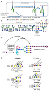
Figure 1
Sialylation pathways in glycoproteins. The protein motifs, sugar symbols, subcellular compartments, and glycosyltransferases involved are shown. (A) Core N-glycosylation in ER [8]. (B) N-glycan sialylation in the Golgi [8,109]. (C) Polysialylation in the Golgi [45]. (D) O-glycan sialylation pathways [8,109].
Figure 2
Figure 2
Sialylation pathways for glycosphingolipids [13,48,59]. Reaction steps and the enzymes involved in the biosynthetic pathways, sugar symbols, as well as subcellular compartments, and the glycosyltransferases involved are shown. According to the nomenclature [110], “G” states the ganglio-series of glycosphingolipids, whereas “A, M, D, T, Q, P” indicate the presence of zero (absent), one (mono-), two (di-), three (tri), four (quadra), and five (penta) sialic acid residues, respectively. Gangliosides with core structure “Galβ1-3GalNAcβ1-4Galβ1-4Glcβ1-1Cer” are indicated with number “1”, those lacking the terminal Gal are labeled with “2”, those lacking the terminal Galβ1-3GalNAc are labeled with “3”, and GM4 is a ganglioside of GalCer. The “0-, a-, b-, c-” series include gangliosides with 0, 1, 2, and 3 sialic acid residues linked to innermost Gal.
Figure 3
Figure 3
Enzymatic specificities for human STs [7]. Four groups and twenty members of human STs are listed. The preferred and alternative products they produce are indicated. Classification A–C based on substrate specificity toward glycoproteins or glycolipids are also designated.
Figure 5
Figure 5
Therapeutic applications with sialylation. (A) Long-acting biotherapeutics with engineered sialylation [338]. Sialylated glycoproteins are not recognized by asialoglycoprotein (ASGP) receptors in the liver and are thus protected from uptake and degradation. (B) Sialic acid-modified micelles or lipid nucleotide particles that bind to E-selectin in tumor tissues [106]. The drug-loaded micelles or mRNA-containing LNPs can be coated with sialic acids for site targeting.

Similar articles

References

    1. Lundblad A. Gunnar Blix and his discovery of sialic acids. Fascinating molecules in glycobiology. Upsala J. Med. Sci. 2015;120:104–112. doi: 10.3109/03009734.2015.1027429. - DOI - PMC - PubMed
    1. Schauer R., Kamerling J.P. Exploration of the Sialic Acid World. Adv. Carbohydr. Chem. Biochem. 2018;75:1–213. - PMC - PubMed
    1. Blix G.S., Svennerholm L., Werner I., Finsnes E., Sorensen J.S., Sorensen N.A. The Isolation of Chondrosamine from Gangliosides and from Submaxillary Mucin. Acta Chem. Scand. 1952;6:358–362. doi: 10.3891/acta.chem.scand.06-0358. - DOI
    1. Blix F.G., Gottschalk A., Klenk E. Proposed nomenclature in the field of neuraminic and sialic acids. Nature. 1957;179:1088. doi: 10.1038/1791088b0. - DOI - PubMed
    1. Schnaar R.L., Gerardy-Schahn R., Hildebrandt H. Sialic acids in the brain: Gangliosides and polysialic acid in nervous system development, stability, disease, and regeneration. Physiol. Rev. 2014;94:461–518. doi: 10.1152/physrev.00033.2013. - DOI - PMC - PubMed

LinkOut - more resources