. 2024 Nov 10;25(22):12065.
doi: 10.3390/ijms252212065.

Human-Brain-Derived Ischemia-Induced Stem Cell Transplantation Is Associated with a Greater Neurological Functional Improvement Compared with Human-Bone Marrow-Derived Mesenchymal Stem Cell Transplantation in Mice After Stroke

Affiliations

Human-Brain-Derived Ischemia-Induced Stem Cell Transplantation Is Associated with a Greater Neurological Functional Improvement Compared with Human-Bone Marrow-Derived Mesenchymal Stem Cell Transplantation in Mice After Stroke

Shuichi Tanada et al. Int J Mol Sci. .

Abstract

The transplantation of injury/ischemia-induced stem cells (iSCs) extracted from post-stroke human brains can improve the neurological functions of mice after stroke. However, the usefulness of iSCs as an alternative stem cell source remains unclear. The current study aimed to assess the efficacy of iSC and mesenchymal stem cell (MSC) transplantation. In this experiment, equal numbers of human brain-derived iSCs (h-iSCs) (5.0 × 10⁴ cells/μL) and human bone marrow-derived MSCs (h-MSCs) (5.0 × 10⁴ cells/μL) were intracranially transplanted into post-stroke mouse brains after middle cerebral artery occlusion. Results showed that not only h-iSC transplantation but also h-MSC transplantation activated endogenous neural stem/progenitor cells (NSPCs) around the grafted sites and promoted neurological functional improvement. However, mice that received h-iSC transplantation experienced improvement in a higher number of behavioral tasks compared with those that received h-MSC transplantation. To investigate the underlying mechanism, NSPCs extracted from the ischemic areas of post-stroke mouse brains were cocultured with h-iSCs or h-MSCs. After coincubation, NSPCs, h-iSCs, and h-MSCs were selectively collected via fluorescence-activated cell sorting. Next, their traits were analyzed via microarray analysis. The genes related to various neuronal lineages in NSPCs after coincubation with h-iSCs were enriched compared with those in NSPCs after coincubation with h-MSCs. In addition, the gene expression patterns of h-iSCs relative to those of h-MSCs showed that the expression of genes related to synapse formation and neurotransmitter-producing neurons increased more after coincubation with NSPCs. Hence, cell-cell interactions with NSPCs promoted transdifferentiation toward functional neurons predominantly in h-iSCs. In accordance with these findings, immunohistochemistry showed that the number of neuronal networks between NSPCs and h-iSCs was higher than that between NSPCs and h-MSCs. Therefore, compared with h-MSC transplantation, h-iSC transplantation is associated with a higher neurological functional improvement, presumably by more effectively modulating the fates of endogenous NSPCs and grafted h-iSCs themselves.

Keywords: cell transplantation; injury/ischemia-induced stem cells; ischemic stroke; mesenchymal stem cells; neural stem/progenitor cells; neurological function.


Conflict of interest statement

The Department of Therapeutic Progress in Brain Diseases is financially supported by Nippon Zoki Pharmaceutical Co., Ltd. and CLEA Japan, Inc. The sponsors had no role in the work, including study design, data collection, data analysis, data interpretation, and manuscript writing.

Figures

Figure 1
Figure 1
(A,B) Heatmap (A) and scatter plot (B) analyses of h-iSCs and h-MSCs. (C,D) h-iSCs (C) and h-MSCs (D) were immunostained with nestin (nestin [(C,D): green] and DAPI [(C,D): blue]). (E) scRNA-seq analysis of h-iSCs and h-MSCs. (FJ) h-iSCs (F) or h-MSCs (G) were labeled with mCherry. Then, mCherry+ h-iSCs (H) or h-MSCs (I) were grafted 6 weeks after MCAO (J). (KM) mCherry+ h-iSCs were transplanted around the ischemic areas of nestin-GFP transgenic mice. Immunohistochemistry 3 days after transplantation showed that in addition to the GFP+ cells in the SVZ, several GFP+ NSPCs were located around the grafted mCherry+ h-iSCs (GFP [(L,M): green], mCherry [(L,M): red], and DAPI [L,M: blue]). (NP) mCherry+ h-MSCs were transplanted around the ischemic areas of nestin-GFP transgenic mice. Immunohistochemistry 3 days after transplantation showed that in addition to the GFP+ cells in the SVZ, several GFP+ NSPCs were located around the grafted mCherry+ h-MSCs (GFP [(O,P): green], mCherry [(O,P): red], and DAPI [(O,P): blue]). Scale bars: 50 µm (C,D), 100 µm (FI), 200 µm (L,O), and 50 µm (M,P). Abbreviations: DAPI, 4′,6-diamidino-2-phenylindole; GFP, green fluorescent protein; iSC, injury/ischemia-induced stem cell; MSC, mesenchymal stem cell; MCAO, middle cerebral artery occlusion; NSPC, neural stem/progenitor cell; scRNA-seq, single-cell RNA sequencing; SVZ, subventricular zone.
Figure 2
Figure 2
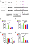
(A) Behavioral tests were performed on the four groups: (1) mice injected with PBS after a sham operation (sham/PBS group, n = 12), (2) mice injected with PBS after MCAO (MCAO/PBS group, n = 8), (3) mice administered h-iSCs after MCAO (MCAO/h-iSC group, n = 12), and (4) mice administered h-MSCs after MCAO (MCAO/h-MSC group, n = 10). (BE) Performance levels in the basket (B), open-field (C), hot plate (D), and open-space swim (E) tests among the four groups. * p < 0.05 between the sham/PBS group (BE). # p < 0.05 between the MCAO/PBS group (BE). Abbreviations: iSC, injury/ischemia-induced stem cell; MCA, middle cerebral artery; MCAO, middle cerebral artery occlusion; MSC, mesenchymal stem cell; PBS, phosphate-buffered saline.
Figure 3
Figure 3
(AC) Regionally derived NSPCs from post-stroke mouse brains were incubated alone (A) or cocultured with GFP+ h-iSCs (B) or GFP+ h-MSCs (C) for immunohistochemistry. (DG) Immunohistochemistry showed that the numbers of nestin+ NSPCs (GFP/nestin+ cells) in samples cocultured with h-iSCs (E) and h-MSCs (F) were significantly higher than those in samples cocultured with NSPCs alone (D). However, the numbers of nestin+ NSPCs in samples cocultured with h-iSCs were significantly higher than those in samples cocultured with h-MSCs (G) (nestin [(DF): red], GFP [(E,F): green], and DAPI [(DF): blue]). Scale bars: 100 µm (DF). * p < 0.05 between the groups (G). n = 3 (12 data points) for each group (G). Abbreviations: DAPI, 4′,6-diamidino-2-phenylindole; GFP, green fluorescent protein; iSC, injury/ischemia-induced stem cell; MSC, mesenchymal stem cell; NSPC, neural stem/progenitor cell.
Figure 4
Figure 4
(A,B) NSPCs were cocultured with h-iSCs (A) or h-MSCs (B) for microarray analysis. (C,D) Scatter plots showing the distribution of genes upregulated more than 5-fold in NSPCs cocultured with h-iSCs relative to NSPCs cocultured with h-MSCs ((C), red plots) or genes upregulated more than 5-fold in NSPCs cocultured with h-MSCs relative to NSPCs cocultured with h-iSCs ((D), green plots). (E,F) List of the top 20 categories for genes overexpressed in NSPCs cocultured with h-iSCs (E) and NSPCs cocultured with h-MSCs (F) based on GO analysis. The former (E) included genes categorized in the “Cell adhesion molecules” category (a red arrow).
Figure 5
Figure 5
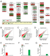
(A) Pathway analysis of the cell lineage map for neuronal differentiation showed that the expression of various neural-lineage-related genes in NSPCs after coincubation with h-iSCs was significantly upregulated (2-fold higher, red box) and/or downregulated (2-fold lower, green box) compared with that in NSPCs after coincubation with h-MSCs. (BD) The scatter plot analysis showed the distribution of significantly upregulated (2-fold higher, red plots) and/or downregulated (2-fold lower, green plots) genes subcategorized into “Immature neuron” and “Mature neuron” (B), “Astrocyte” (C), and “Oligodendrocyte” (D) in the cell lineage map for neuronal differentiation. (EG) The numbers of significantly upregulated (2-fold higher, red box) and/or downregulated (2-fold lower, green box) genes subcategorized as “Immature neuron” and “Mature neuron” (E), “Astrocyte” (F), and “Oligodendrocyte” (G) in the cell lineage map for neuronal differentiation. Abbreviations: iSC, injury/ischemia-induced stem cell; MSC, mesenchymal stem cell; NSPC, neural stem/progenitor cell.
Figure 6
Figure 6
(A,B) h-iSCs were cultured alone (A) or cocultured with NSPCs (B) for microarray analysis. (C) Pathway analysis of the cell lineage map for neuronal differentiation showed that the expression of various neural lineage-related genes in h-iSCs after coincubation with NSPCs was significantly upregulated (2-fold higher, red box) and/or downregulated (2-fold lower, green box) compared with those in h-iSCs alone. (DF) The scatter plot analysis showed the distribution of significantly upregulated (2-fold higher, red plots) and/or downregulated (2-fold lower, green plots) genes subcategorized into “Stem cell” and “Neural progenitor” (D), “Immature neuron” and “Mature neuron” (E), and “Pre-synapse” and “Post-synapse” (F) in the cell lineage map for neuronal differentiation. (GI) The numbers of significantly upregulated (2-fold higher, red box) and/or downregulated (2-fold lower, green box) genes subcategorized into “Stem cell” and “Neural progenitor” (G), “Immature neuron” and “Mature neuron” (H), and “Pre-synapse” and “Post-synapse” (I) in the cell lineage map for neuronal differentiation. Abbreviations: iSC, injury/ischemia-induced stem cell; NSPC, neural stem/progenitor cell.
Figure 7
Figure 7
(A,B) h-MSCs were cultured alone (A) or cocultured with NSPCs (B) for microarray analysis. (C) Pathway analysis of the cell lineage map for neuronal differentiation showed that the expression of various neural lineage-related genes in h-MSCs after coincubation with NSPCs was significantly upregulated (2-fold higher, red box) and/or downregulated (2-fold lower, green box) compared with that in h-MSCs alone. (DF) The scatter plot analysis showed the distribution of significantly upregulated (2-fold higher, red plots) and/or downregulated (2-fold lower, green plots) genes subcategorized into “Stem cell” and “Neural progenitor” (D), “Immature neuron” and “Mature neuron” (E), and “Pre-synapse” and “Post-synapse” (F) in the cell lineage map for neuronal differentiation. (GI) The numbers of significantly upregulated (2-fold higher, red box) and/or downregulated (2-fold lower, green box) genes subcategorized into “Stem cell” and “Neural progenitor” (G), “Immature neuron” and “Mature neuron” (H), and “Pre-synapse” and “Post-synapse” (I) in the cell lineage map for neuronal differentiation. Abbreviations: MSC, mesenchymal stem cell; NSPC, neural stem/progenitor cell.
Figure 8
Figure 8
(A,B) Pathway analysis of the cell lineage map for neuronal differentiation was performed between h-iSCs and h-MSCs before (A) and after coincubation with NSPCs (B). The expression of neural lineage-related genes in h-iSCs relative to those in h-MSCs was significantly upregulated (2-fold higher, red box) and/or downregulated (2-fold lower, green box). Abbreviations: iSC, injury/ischemia-induced stem cells; MSC, mesenchymal stem cell; NSPC, neural stem/progenitor cell.
Figure 9
Figure 9
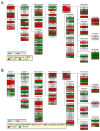
(AL) Pathway analysis of the cell lineage map for neuronal differentiation was performed between h-iSCs and h-MSCs before and after coincubation with NSPCs. The expression of significantly upregulated (2-fold higher, red box) and/or downregulated (2-fold lower, green box) genes in h-iSCs relative to those in h-MSCs, which were subcategorized into “Neural progenitor” (A), “Immature neuron” (B), “Mature neuron” (C), “Pre-synapse” (D), “Post-synapse” (E), “Glutamatergic” (F), “Glycinergic” (G), “Dopaminergic” (H), “Noradrenergic” (I), “GABAergic” (J), “Cholinergic” (K), and “Serotonergic” (L) in the cell lineage map for neuronal differentiation, are presented. Abbreviations: iSC, injury/ischemia-induced stem cell; MSC, mesenchymal stem cell; NSPC, neural stem/progenitor cell.
Figure 10
Figure 10
(A,B) GFP+ h-iSCs (A) or h-MSCs (B) were cocultured with NSPC-derived neurospheres for 2 weeks. (C,D) Immunocytochemistry of NSPC-derived neurons (GFP/MAP2+ cells) cocultured with h-iSCs (C) and h-MSCs (D) (MAP2 [(C,D): red], GFP [(C,D): green], and DAPI [(C,D): blue]). (E) The ratio of NSPC-derived neurons (GFP/MAP2+ cells to GFP/DAPI+ cells) after coculture with h-iSCs was higher than that after coculture with h-MSCs. (F) The ratio of h-iSC-derived neurons (GFP+/MAP2+ cells to GFP+/DAPI+ cells) was significantly higher than that of h-MSC-derived neurons (GFP+/MAP2+ cells to GFP+/DAPI+ cells) in the presence of NSPCs. (G) The number of NSPC-derived neurons (GFP/MAP2+ cells) that interacted with h-iSC-derived neurons (GFP+/MAP2+ cells) was significantly higher than that of NSPC-derived neurons that interacted with h-MSC-derived neurons (GFP+/MAP2+ cells). Scale bars: 100 µm (C,D). * p < 0.05 between the h-iSC(+) and h-MSC(+) groups (E,G). p < 0.05 between the h-iSC and h-MSC groups (F). n = 3 (nine data points) for each group (EG). Abbreviations: DAPI, 4′,6-diamidino-2-phenylindole; GFP, green fluorescent protein; iSC, injury/ischemia-induced stem cells; NSPC, neural stem/progenitor cell; MAP2, microtubule-associated protein 2; MSC, mesenchymal stem cell.

Similar articles

References

    1. Doi D., Samata B., Katsukawa M., Kikuchi T., Morizane A., Ono Y., Sekiguchi K., Nakagawa M., Parmar M., Takahashi J. Isolation of human induced pluripotent stem cell-derived dopaminergic progenitors by cell sorting for successful transplantation. Stem Cell Rep. 2014;2:337–350. doi: 10.1016/j.stemcr.2014.01.013. - DOI - PMC - PubMed
    1. Hamblin M.H., Murad R., Yin J., Vallim G., Lee J.P. Modulation of gene expression on a transcriptome-wide level following human neural stem cell transplantation in aged mouse stroke brains. Exp. Neurol. 2022;347:113913. doi: 10.1016/j.expneurol.2021.113913. - DOI - PMC - PubMed
    1. Vats K., Sarmah D., Datta A., Saraf J., Kaur H., Pravalika K., Wanve M., Kalia K., Borah A., Dave K.R., et al. Intra-arterial Stem Cell Therapy Diminishes Inflammasome Activation After Ischemic Stroke: A Possible Role of Acid Sensing Ion Channel 1a. J. Mol. Neurosci. 2021;71:419–426. doi: 10.1007/s12031-019-01460-3. - DOI - PubMed
    1. Kikuchi-Taura A., Okinaka Y., Takeuchi Y., Ogawa Y., Maeda M., Kataoka Y., Yasui T., Kimura T., Gul S., Claussen C., et al. Bone Marrow Mononuclear Cells Activate Angiogenesis via Gap Junction-Mediated Cell-Cell Interaction. Stroke. 2020;51:1279–1289. doi: 10.1161/STROKEAHA.119.028072. - DOI - PubMed
    1. Nakagomi T., Taguchi A., Fujimori Y., Saino O., Nakano-Doi A., Kubo S., Gotoh A., Soma T., Yoshikawa H., Nishizaki T., et al. Isolation and characterization of neural stem/progenitor cells from post-stroke cerebral cortex in mice. Eur. J. Neurosci. 2009;29:1842–1852. doi: 10.1111/j.1460-9568.2009.06732.x. - DOI - PubMed

LinkOut - more resources