Review
. 2024 Nov 10;25(22):12078.
doi: 10.3390/ijms252212078.

Unraveling Macrophage Polarization: Functions, Mechanisms, and "Double-Edged Sword" Roles in Host Antiviral Immune Responses

Affiliations
Review

Unraveling Macrophage Polarization: Functions, Mechanisms, and "Double-Edged Sword" Roles in Host Antiviral Immune Responses

Meng Yao et al. Int J Mol Sci. .

Abstract

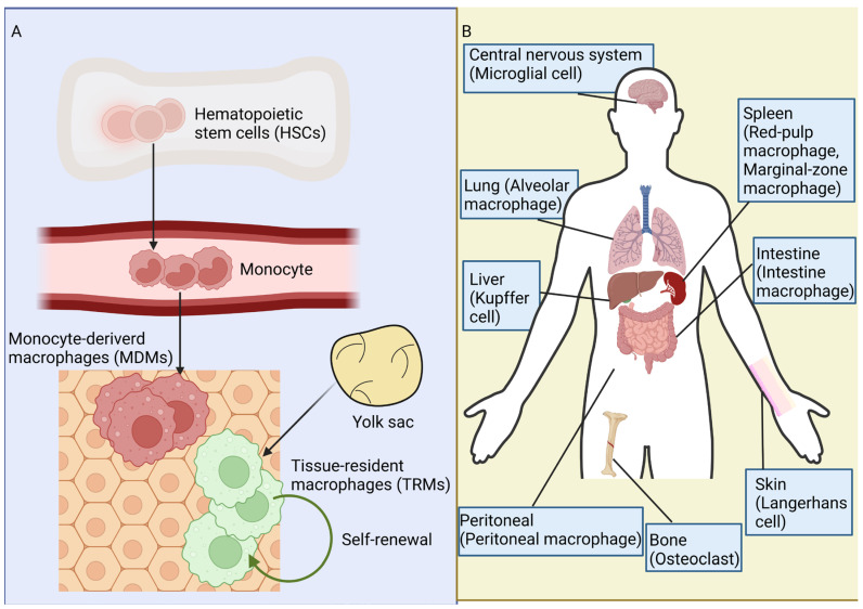
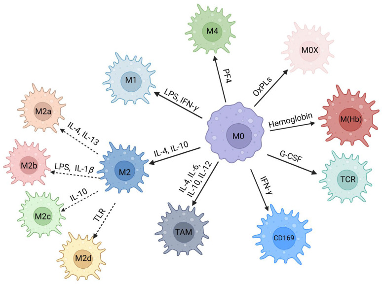
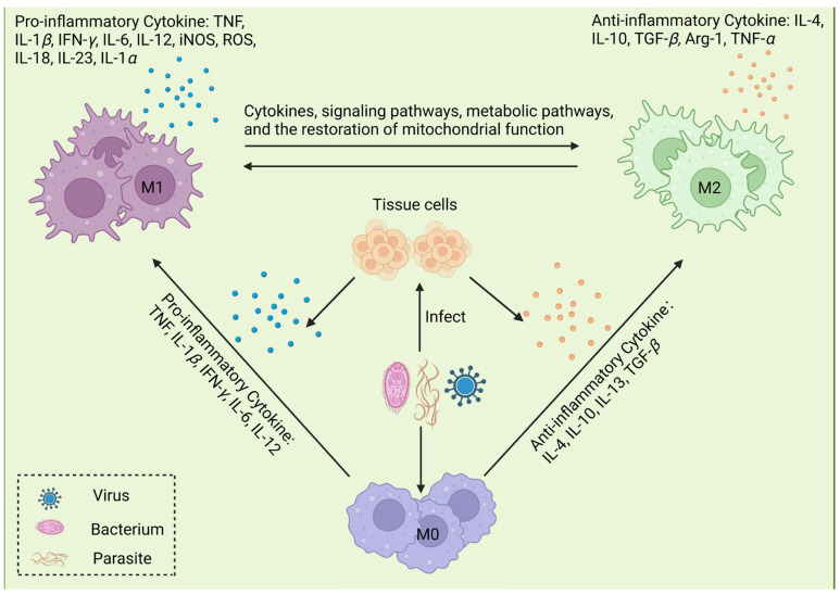
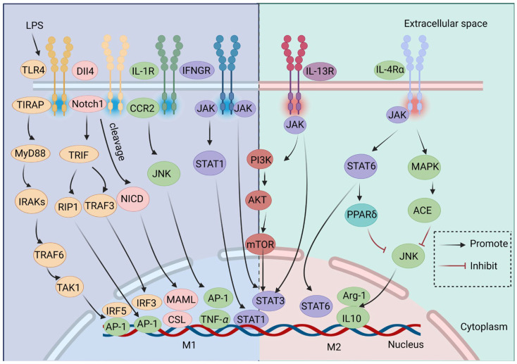
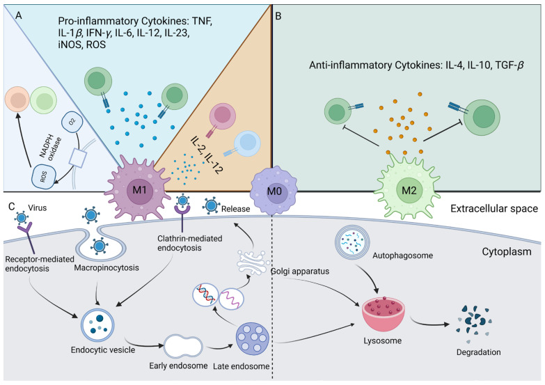
Numerous viruses that propagate through the respiratory tract may be initially engulfed by macrophages (Mφs) within the alveoli, where they complete their first replication cycle and subsequently infect the adjacent epithelial cells. This process can lead to significant pathological damage to tissues and organs, leading to various diseases. As essential components in host antiviral immune systems, Mφs can be polarized into pro-inflammatory M1 Mφs or anti-inflammatory M2 Mφs, a process involving multiple signaling pathways and molecular mechanisms that yield diverse phenotypic and functional features in response to various stimuli. In general, when infected by a virus, M1 macrophages secrete pro-inflammatory cytokines to play an antiviral role, while M2 macrophages play an anti-inflammatory role to promote the replication of the virus. However, recent studies have shown that some viruses may exhibit the opposite trend. Viruses have evolved various strategies to disrupt Mφ polarization for efficient replication and transmission. Notably, various factors, such as mechanical softness, the altered pH value of the endolysosomal system, and the homeostasis between M1/M2 Mφs populations, contribute to crucial events in the viral replication cycle. Here, we summarize the regulation of Mφ polarization, virus-induced alterations in Mφ polarization, and the antiviral mechanisms associated with these changes. Collectively, this review provides insights into recent advances regarding Mφ polarization in host antiviral immune responses, which will contribute to the development of precise prevention strategies as well as management approaches to disease incidence and transmission.

Keywords: antiviral immunity; immune escape; macrophage polarization; macrophages; viruses.


Conflict of interest statement

The authors declare that the research was conducted in the absence of any commercial or financial relationships that could be construed as potential conflicts of interest.

Figures

Figure 1
Figure 1
Origin and distribution of macrophages (Mφs). (A) The origin of Mφs. Hematopoietic stem cells (HSCs) in the bone marrow can differentiate into monocytes, which subsequently enter the blood and mature. These circulating monocytes are capable of migrating through blood vessels into tissue, where they differentiate into monocyte-derived Mφs (MDMs). Tissue-resident macrophages (TRMs) are mainly generated by the self-renewal and regeneration of existing Mφs. Many TRMs are established during embryonic development rather than adult blood monocytes, and they operate independently of one another. (B) The distribution of Mφs in different organs. Upon migrating to specific tissues, Mφs differentiate into tissue-specific Mφs. These include microglia in the central nervous system, Kupffer cells in the liver, Langerhans cells in the epidermis, osteoclasts within the skeletal system, alveolar Mφs in the lung, histocytes in connective tissues, and metallophilic cells in the spleen.
Figure 2
Figure 2
Classification of macrophage (Mφ) polarization. Under stimulation by different factors, M0 Mφs can be polarized into different phenotypes. M1 Mφs are classically activated in response to lipopolysaccharides (LPSs) and interferon gamma (IFN-γ). Conversely, upon stimulation by interleukin 4 (IL-4) and IL-10, M2 Mφs are activated instead, which can be further divided into M2a, M2b, M2c, or M2d phenotypes depending on the specific stimuli. In the presence of IFN-γ and granulocyte colony-stimulating factor (G-CSF), Mφs may be polarized into CD169+ Mφs and TCR+ Mφs, respectively. Under certain conditions, such as within the tumor microenvironment, tumor-associated Mφs are polarized. In atherosclerosis, they are polarized into M4, Mox, and M(Hb) phenotypes by platelet factor 4 (PF4), oxidized phospholipids (OxPLs), and hemoglobin. TLR—toll-like receptor.
Figure 3
Figure 3
Mechanisms of microorganism-induced macrophage (Mφ) polarization. Following invasion of the body by pathogenic microorganisms (bacteria, viruses, parasites, etc.), these pathogens infect corresponding tissue cells, which subsequently secrete a series of cytokines, such as tumor necrosis factor-alpha (TNF-α), TNF-β, interleukin-6 (IL-6), IL-12, IL-1β, IL-4, and IL-10. With the secretion of cytokines, Mφs are recruited to the injury site, where they are polarized into different phenotypes under stimulation by different cytokines, thus participating in the immune response and maintaining body homeostasis. M1 Mφs secrete pro-inflammatory cytokines, including TNF, IL-1β, interferon gamma (IFN-γ), IL-6, IL-12, inducible nitric oxide synthase (iNOS), and reactive oxygen species (ROS), which participate in the early inflammatory response, while M2 Mφs can release IL-10, TGF-β, and other anti-inflammatory cytokines.
Figure 4
Figure 4
Signaling pathways regulating macrophage (Mφ) polarization. Distinct transcription factors and signaling pathways influence the polarization of M1 and M2 Mφs. The JAK/STAT, PI3K/Akt, JNK, Notch and TLR signaling pathways serve as primary examples of these regulatory mechanisms. Interferon gamma (IFN-γ) binds to its receptor, interferon gamma receptor (IFNGR), leading to the polarization of Mφs toward the M1 phenotype by phosphorylating STAT1 and activating the JAK/STAT1 signaling pathway. This process also initiates the transcription of IFN-stimulated genes. The binding of interleukin 4 (IL-4) or IL-13 to their respective receptors results in the activation of STAT3 or STAT6, respectively; this promotes the transcription of anti-inflammatory cytokines that drive Mφs toward an anti-inflammatory M2 phenotype. Additionally, upon interaction with its receptor, the Notch protein is activated and the Notch intracellular domain (NICD) is translocated to the nucleus. This event subsequently enhances the production of pro-inflammatory cytokines and M1-related encoding genes like mastermind-like transcriptional coactivator (MAML) and CBF1/Su(H)/LAG-1 (CSL). There are no precise limits to the PI3K/Akt/mTOR signaling pathway’s ability to control M1 and M2 Mφs. IL-1 and IL-4 activate the JNK signaling pathway, which causes Mφ polarization toward the M1 and M2 phenotypes, respectively. The MyD88-independent pathway of the TLR4 signaling pathway mainly activates interferon regulatory factor 3 (IRF3) to induce type I IFNs. DLL4, delta-like ligand 4; CRR2, C-C motif chemokine receptor 2; JNK, c-Jun N-terminal kinase; JAK, Janus kinase; STAT, signal transducer and activator of transcription; MAPK, mitogen-activated protein kinase; mTOR, mammalian target of rapamycin; Arg-1, arginase 1; AP-1, activator protein 1; ACE, angiotensin-converting enzyme.
Figure 5
Figure 5
Dual identity of macrophage (Mφ) polarization in antiviral immunity. (A) Antiviral mechanisms of M1 Mφs. During the process of viral infections, ROS produced by M1 Mφs can create a superoxide environment that stimulates the immune system and plays positively regulatory roles in virus clearance. M1 Mφs possess the ability to secrete pro-inflammatory cytokines, including IL-1-β, TNF-β, IFN-γ, IL-6, IL-12, IL-23, iNOS, ROS, etc. To further block the invasion of the virus, M1 Mφs can also activate additional immune cells to fight against the virus. (B) Immunosuppressive effects of M2 Mφs. M2 Mφs secrete anti-inflammatory cytokines, like TGF-β and IL-10. M2 Mφs, by controlling the activation of T cells, can induce immunosuppression and promote tumorigenesis while inhibiting the production of anti-tumor immune responses by T cells. (C) The endosomal–lysosomal system in M1 and M2 Mφs. Viruses primarily enter Mφs through endocytosis, including macropinocytosis, receptor-mediated endocytosis, and clathrin-mediated endocytosis (CME). Upon entry into the cell, the virus undergoes endocytosis, progressing through early and late endosomes before being degraded within the lysosomes. The acidic environment of endosomes and alkaline nature of lysosomes in M1 Mφs facilitate viral replication, leading to eventual viral escape. Conversely, the acidic lysosomes of M2 Mφs possess the ability to degrade the virus, thereby preventing its spread. ROS, reactive oxygen species; iNOS, inducible nitric oxide synthase.

Similar articles

References

    1. Chen S., Saeed A.F.U.H., Liu Q., Jiang Q., Xu H., Xiao G.G., Rao L., Duo Y. Macrophages in immunoregulation and therapeutics. Signal Transduct. Target Ther. 2023;8:207. doi: 10.1038/s41392-023-01452-1. - DOI - PMC - PubMed
    1. Bian Z., Gong Y., Huang T., Lee C.Z.W., Bian L., Bai Z., Shi H., Zeng Y., Liu C., He J., et al. Deciphering human macrophage development at single-cell resolution. Nature. 2020;582:571–576. doi: 10.1038/s41586-020-2316-7. - DOI - PubMed
    1. Ammarrah U., Pereira-Nunes A., Delfini M., Mazzone M. From monocyte-derived macrophages to resident macrophages-how metabolism leads their way in cancer. Mol. Oncol. 2024;18:1739–1758. doi: 10.1002/1878-0261.13618. - DOI - PMC - PubMed
    1. Van Breedam W., Delputte P.L., Van Gorp H., Misinzo G., Vanderheijden N., Duan X., Nauwynck H.J. Porcine reproductive and respiratory syndrome virus entry into the porcine macrophage. J. Gen. Virol. 2010;91:1659–1667. doi: 10.1099/vir.0.020503-0. - DOI - PubMed
    1. Chen D., Xie J., Fiskesund R., Dong W., Liang X., Lv J., Jin X., Liu J., Mo S., Zhang T., et al. Chloroquine modulates antitumor immune response by resetting tumor-associated macrophages toward M1 phenotype. Nat. Commun. 2018;9:873. doi: 10.1038/s41467-018-03225-9. - DOI - PMC - PubMed

LinkOut - more resources