Review
. 2024 Nov 11;25(22):12082.
doi: 10.3390/ijms252212082.

Innate Immunity and Synovitis: Key Players in Osteoarthritis Progression

Affiliations
Review

Innate Immunity and Synovitis: Key Players in Osteoarthritis Progression

Veronica Panichi et al. Int J Mol Sci. .

Abstract

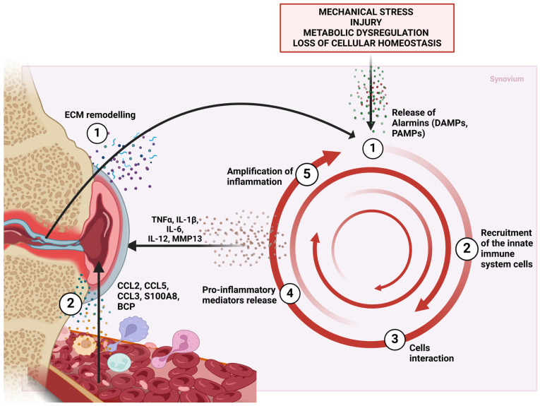
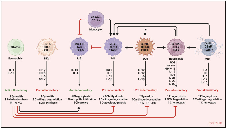
Osteoarthritis (OA) is a chronic progressive disease of the joint. Although representing the most frequent cause of disability in the elderly, OA remains partly obscure in its pathogenic mechanisms and is still the orphan of resolutive therapies. The concept of what was once considered a "wear and tear" of articular cartilage is now that of an inflammation-related disease that affects over time the whole joint. The attention is increasingly focused on the synovium. Even from the earliest clinical stages, synovial inflammation (or synovitis) is a crucial factor involved in OA progression and a major player in pain onset. The release of inflammatory molecules in the synovium mediates disease progression and worsening of clinical features. The activation of synovial tissue-resident cells recalls innate immunity cells from the bloodstream, creating a proinflammatory milieu that fuels and maintains a damaging condition of low-grade inflammation in the joint. In such a context, cellular and molecular inflammatory behaviors in the synovium could be the primum movens of the structural and functional alterations of the whole joint. This paper focuses on and discusses the involvement of innate immunity cells in synovitis and their role in the progression of OA.

Keywords: inflammaging; inflammation; innate immunity; innate immunity cells; loss of chondrocyte homeostasis; low-grade inflammation; osteoarthritis; osteoarthritis pathogenesis; synovitis; synovium.


Conflict of interest statement

The authors declare no conflicts of interest.

Figures

Figure 1
Figure 1
Cellular mechanisms involved in OA synovitis in osteoarthritis. OA is a multifactorial disease characterized by several risk factors such as mechanical stress, injury, metabolic dysregulation, but, above all, loss of chondrocyte cellular homeostasis that affects all other cells resident in the joint tissues. Deregulation at the cellular level triggers inflammatory mechanisms and determines the establishment of a self-sustaining low-grade inflammation, also known as “inflammaging”. The triggering of synovitis in OA is not entirely clear due to the overlap and mutual dependence of the mechanisms involved. The most accepted theory is that the loss of chondrocyte homeostasis is the first step in the chain of events. Chondrocytes are the only cell type of articular cartilage. Activation of inflammation-related signaling pathways induces the release of cartilage matrix-degrading enzymes such as MMP-13 and MMP-3, responsible for ECM remodeling in articular cartilage. ECM fragments (through the DAMPs they expose and the subsequent interaction of DAMPs with PRRs of immune cells) and proinflammatory mediators (such as cytokines) stimulate the inflammatory response of the synovium (1). Activation of inflammation leads to the recruitment of innate immune cells from the bloodstream (2) along with the activation of immunocompetent cells that reside in the synovium (mainly macrophages). The interaction between different immune cells in the synovium (3) fuels the production of multiple proinflammatory factors (4) that cause further tissue damage (5). This endless cycle is pivotal in sustaining the inflammatory milieu leading to the progression of the OA. Abbreviations: DAMPs, danger-associated molecular patterns; PAMPs, pathogen-associated molecular patterns; PRRs, pattern recognition receptors; CCL, C-C motif chemokine ligand; IL, interleukin; TNF-α, tumor necrosis factor-α; MMP, matrix metalloprotease.
Figure 2
Figure 2
Cells of innate immunity and network of mediators in OA synovitis. Synovitis in osteoarthritis is mainly driven by the activation of innate immunity. This activation is triggered by proinflammatory factors released following the loss of cellular homeostasis. Each type of immunocompetent cell releases mediators to promote/counteract low-grade inflammation. Eosinophils and M2 macrophages exert an anti-inflammatory activity under the modulation of the JAK/STAT signaling pathway and the release of anti-inflammatory cytokines (IL-4, IL-10, IL-13). Thus, boosting these immune cells and the release of their mediators could be useful to re-establish cellular homeostasis and counteract synovitis. In contrast, M1 macrophages, NK cells, DCs, neutrophils, and MCs actively fuel the vicious cycle that keeps low-grade inflammation and synovitis alive, leading to OA progression. The release of proinflammatory mediators (such as IL-1, TNF-α, and ROS) following immune cell activation leads to worsening of synovitis features, cartilage degradation, and bone remodeling. Furthermore, the chemotactic activity of these molecules attracts more immunocompetent cells from the bloodstream, thus corroborating the proinflammatory milieu. Abbreviations: IL, interleukin; CD, cluster of differentiation; NKs, natural killer cells; M2, anti-inflammatory macrophages; M1, proinflammatory macrophages; DCs, dendritic cells; MCs, mast cells; Th, T helper; JAK/STAT, Janus kinase/signal transducer and activator of transcription; MHC, major histocompatibility complex; NF-κB, Nuclear factor kappa-light-chain-enhancer of activated B cells; TLR-2/4, Toll-like receptor 2/4; GNLY, Granulysin; PAR-2, protease-activated receptor 2; FcεRI, high-affinity IgE receptor; C5aR, complement receptor C5a; ROS, reactive oxygen species; MCP-1, Monocyte Chemotactic Protein 1; MMP-13, metalloprotease 13; ECM, extracellular matrix.

Similar articles

Cited by

References

    1. Long H., Liu Q., Yin H., Wang K., Diao N., Zhang Y., Lin J., Guo A. Prevalence Trends of Site-Specific Osteoarthritis From 1990 to 2019: Findings From the Global Burden of Disease Study 2019. Arthritis Rheumatol. 2022;74:1172–1183. doi: 10.1002/art.42089. - DOI - PMC - PubMed
    1. Wong A.Y., Samartzis D., Maher C. The global burden of osteoarthritis: Past and future perspectives. Lancet Rheumatol. 2023;5:e496–e497. doi: 10.1016/S2665-9913(23)00207-2. - DOI - PubMed
    1. Nelson A.E. Multiple joint osteoarthritis (mjoa): What’s in a name? Osteoarthr. Cartil. 2024;32:234–240. doi: 10.1016/j.joca.2023.10.008. - DOI - PMC - PubMed
    1. Palazzo C., Nguyen C., Lefevre-Colau M.-M., Rannou F., Poiraudeau S. Risk factors and burden of osteoarthritis. Ann. Phys. Rehabil. Med. 2016;59:134–138. doi: 10.1016/j.rehab.2016.01.006. - DOI - PubMed
    1. Dorio M., Deveza L.A. Phenotypes in osteoarthritis: Why do we need them and where are we at? Clin. Geriatr. Med. 2022;38:273–286. doi: 10.1016/j.cger.2021.11.002. - DOI - PubMed

LinkOut - more resources