. 2024 Nov 11;25(22):12090.
doi: 10.3390/ijms252212090.

An Immune-Independent Mode of Action of Tacrolimus in Promoting Human Extravillous Trophoblast Migration Involves Intracellular Calcium Release and F-Actin Cytoskeletal Reorganization

Affiliations

An Immune-Independent Mode of Action of Tacrolimus in Promoting Human Extravillous Trophoblast Migration Involves Intracellular Calcium Release and F-Actin Cytoskeletal Reorganization

Ahmad J H Albaghdadi et al. Int J Mol Sci. .

Abstract

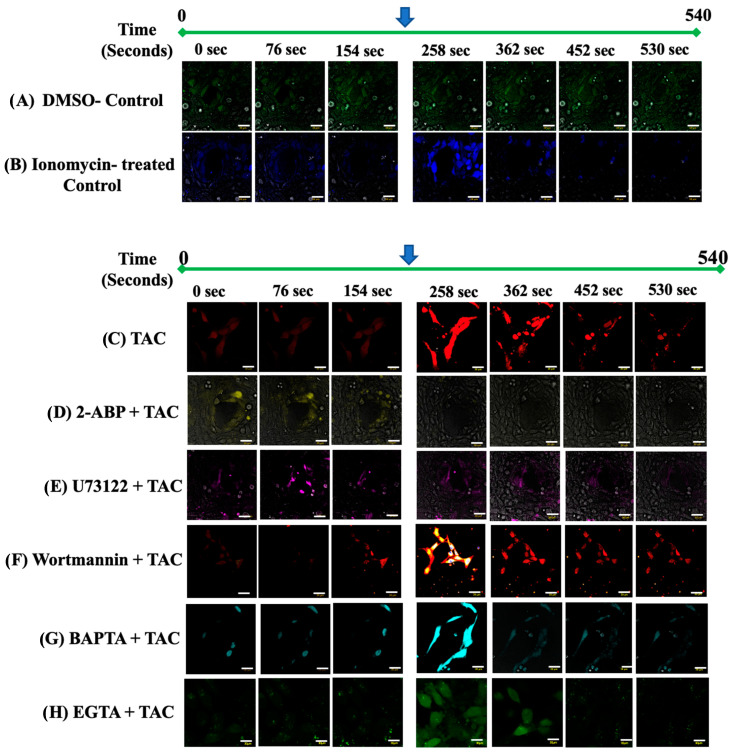
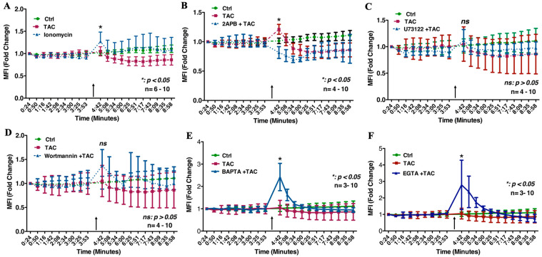
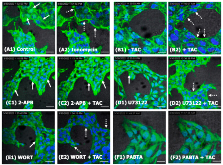
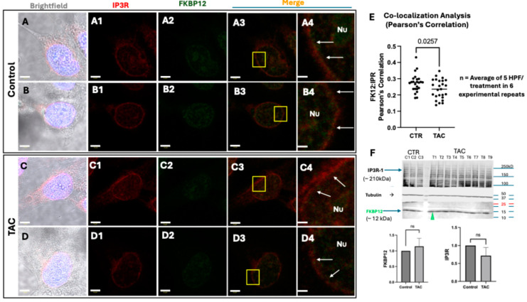
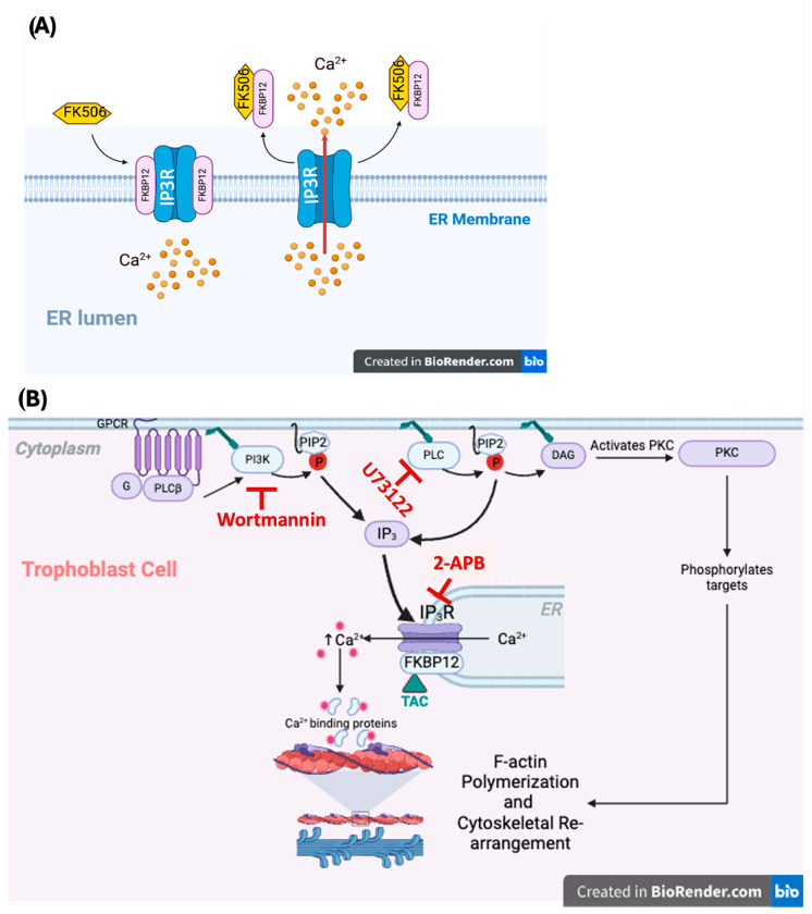
We have previously reported that the calcineurin inhibitor macrolide immunosuppressant Tacrolimus (TAC, FK506) can promote the migration and invasion of the human-derived extravillous trophoblast cells conducive to preventing implantation failure in immune-complicated gestations manifesting recurrent implantation failure. Although the exact mode of action of TAC in promoting implantation has yet to be elucidated, the integral association of its binding protein FKBP12 with the inositol triphosphate receptor (IP3R) regulated intracellular calcium [Ca2+]i channels in the endoplasmic reticulum (ER), suggesting that TAC can mediate its action through ER release of [Ca2+]i. Using the immortalized human-derived first-trimester extravillous trophoblast cells HTR8/SVneo, our data indicated that TAC can increase [Ca2+]I, as measured by fluorescent live-cell imaging using Fluo-4. Concomitantly, the treatment of HTR8/SVneo with TAC resulted in a major dynamic reorganization in the actin cytoskeleton, favoring a predominant distribution of cortical F-actin networks in these trophoblasts. Notably, the findings that TAC was unable to recover [Ca2+]i in the presence of the IP3R inhibitor 2-APB indicate that this receptor may play a crucial role in the mechanism of action of TAC. Taken together, our results suggest that TAC has the potential to influence trophoblast migration through downstream [Ca2+]i-mediated intracellular events and mechanisms involved in trophoblast migration, such as F-actin redistribution. Further research into the mono-therapeutic use of TAC in promoting trophoblast growth and differentiation in clinical settings of assisted reproduction is warranted.

Keywords: F-actin cytoskeleton; FK506); FKBP12; [Ca2+]i; extravillous trophoblasts; inositol triphosphate receptor (IP3R); tacrolimus (TAC.


Conflict of interest statement

The authors declare no conflict of interest.

Figures

Figure 1
Figure 1
Tacrolimus spiked [Ca2+]i release in live HTR8/Svneo cells. (AH): Representative photomicrographs of individual live HTR8/SVneo cells visualized by intravital confocal microscopy depicting the intracellular contents of Ca2+ in DMSO-treated control ((A): green), ionomycin ((B): blue), TAC-treated ((C): red), 2-APB + TAC ((D): yellow), U73122 + TAC ((E): purple), Wortmannin + TAC ((F): orange), BAPTA + TAC ((G): turquoise) and EGTA + TAC ((H): green), respectively. The timeframe for Ca2+ imaging was for a total of 540 s. [Ca2+]i release was spiked at the end of minute 3 of live-image tracing (depicted by the blue arrow) by the addition of ionomycin (B), or TAC alone (C) or in the presence of other inhibitors of [Ca2+]i release and Ca2+ chelators (DG). Compared to the DMSO-treated (A) and Ionomycin-treated controls (B), the single use of TAC (10 ng/mL) resulted in a significant increase in [Ca2+]i in the HTR8/SVneo cells (compare the intensity of red color in (C) before and after the addition of TAC). The inability of TAC to spike [Ca2+]i-release in the presence of the IP3R antagonist 2-APB (D) suggests a crucial role for IP3R in mediating the [Ca2+]i modulatory actions of TAC. Unexpectedly, the TAC-induced [Ca2+]i-release in HTR8/SVneo cells was unaffected by inhibitory actions of the PLC inhibitor U73122 (E) as well as the potent and specific phosphatidylinositol 3-kinase (PI3-K) inhibitor Wortmannin (F). The intracellular source of the TAC-induced [Ca2+]i-release is confirmed by the use of intracellular [Ca2+]i chelator BPATA (G) and EGTA (H). Scale bars: (AH) 30 µm; [Ca2+]i is depicted in pseudocolors in (AH).
Figure 2
Figure 2
Real-time tracing of [Ca2+]i-release in live HTR8/SVneo cells. (AF): Fold change in Mean Fluorescence Intensity (MFI) of Fluo-4 as a measurement of [Ca2+]i-release in HTR8/SVneo cells receiving TAC (A) in the presence and absence of the [Ca2+]i-release inhibitors (2-APB: (B), U73122: (C), Wortmannin: (D)), as well as the Ca2+chelators BAPTA (E), and EGTA (F). (A) depicts the influence of TAC administration on spiking [Ca2+]i-release (black arrow) in a comparable intensity to that of the calcium ionophore Ionomycin. (B) depicts pre-incubation with the IP3R inhibitor 2-APB significantly suppressed TAC-induced [Ca2+]i-release in HTR8/SVneo cells (p < 0.05). (C,D): Unlike the suppressive effects of the 2-APB on TAC-induced [Ca2+]i-release, the inhibition of the phospholipase C (PLC) or the phosphatidylinositol 3-kinase (PI3K) by use of the compound U73122 (C) and Wortmannin (D) did not restrict this cellular action of TAC. In (E,F), the spiked TAC-induced [Ca2+]i-release in HTR8/SVneo cells concomitantly treated with the intracellular and extracellular calcium chelators BAPTA (E) and EGTA (F) suggests the release of [Ca2+]i is from the intracellular stores, namely the endoplasmic reticulum. In (AF), HTR8/SVneo cells were pre-incubated for 10 min with the [Ca2+]i-release inhibitors prior to the administration of TAC. Real-time tracing of [Ca2+]i-release in Ionomycin-treated HTR8/SVneo cells was included in (A) for a comparison. Black arrows in (AF) depict spiked [Ca2+]i-release. The time recording was 10 min.
Figure 3
Figure 3
Tacrolimus influences F-actin cytoskeletal re-arrangement in the human-derived first-trimester extravillous trophoblast cells. (AC): Single cell confocal images of control (A) and TAC-treated HTR8/SVneo cells (B,C) labeled with the CellMask GreenTM Actin tracking stain. F-actin is mostly distributed in the form of stress fibers running across the cell body of untreated cells (white arrows in (A1,A2)). 10 min pre-incubation with TAC resulted in a global reorganization of the F-actin filaments manifested in the formation of cortical fibers (white arrows in (B1,B2)). Notably, filopodia-like structures (white arrows in (C1,C2)) were observed among TAC-treated HTR8/SVneo cells evidently demonstrating a tangible outcome of the influence of TAC on F-actin cytoskeletal reorganization suggestive of cell motility. Green: CellMask GreenTM-labled F-actin, Blue: DAPI-stained nuclei. Scale bars: (AC) 10 µm. Nuclei were counterstained with DAPI in (AC).
Figure 4
Figure 4
The influence of [Ca2+]i-release inhibitors and chelators on the structural distribution of F-actin in TAC-treated HTR8/SVneo cells. (AF): Time-dependent cytoskeletal reorganization of F-actin in the HTR8/SVneo cells in response to Ionomycin (A1,A2), TAC (B1,B2), 2-APB (C1,C2), U73122 (D1,D2), Wortmannin (E1,E2) and PABTA (F1,F2), respectively. Note the characteristic distribution of the stress fibers throughout the cytoplasm (solid white arrows) versus the peripherally condensed cortical fiber (dashed white arrows). Failure of F-actin cellular reorganization in the 2-APB-inhibited cells ((C1) vs. (C2)) indicates the dependence of TAC actions on the functional IP3R-signaling pathway. Distinctly, unlike pre-incubation with U73122 (D1,D2) and Wortmannin (E1,E2), the presence of 2-APB (C1,C2) and PABTA (F1,F2) compromised the structural integrity and consequently the visualization of the F-actin cytoskeleton in HTR8/SVneo cells. The recording after the addition of TAC to the inhibitor pre-treated cells was 6 min. Green: CellMask GreenTM-labled F-actin, Blue: DAPI-stained nuclei. Scale bars: (A1F2) 45 µm.
Figure 5
Figure 5
Tacrolimus negatively influenced the co-localization of IP3R and FKBP12 in HTR8/SVneo cells. (AE): Representative confocal images for the immunofluorescent detection of IP3R-1 ((A1D1): red *) and FKBP12 ((A2D2): green *) and their co-localization ((A3D4); firey orange, and co-localization analysis bar graphs in (E)) in PFA-fixed monolayers of control (DMSO-treated) and TAC-treated HTR8/SVneo cells depicting a significant reduction (p = 0.025) in the Pearson’s correlation coefficient of mean fluorescent intensities (MFI) of the co-localization of these two protein components of the ER [Ca2+]i-release channels after 1 h of exposure to TAC (E). Indeed, Pearson’s correlation quantification revealed that IP3R-I and FKBP12 are co-expressed in the same pixel in control cells more than in TAC-treated cells **. Note that the characteristic perinuclear distribution of these two proteins in HTR8/SVeno cells (white arrows in (A4D4), respectively). (F): representative Western blot (Wb) detection of IP3R and FKBP12 in control (experimental repeats C1–C3 in lanes C1–C3) and TAC-treated (experimental repeats T1–T9 in lanes T1–T9) HTR8/SVneo cells, revealing a preservative effect of TAC in maintaining the levels of these two protein components of the ER [Ca2+]i-release channels in trophoblasts as measured by the lack of a significant fold change in their protein band intensities compared to untreated control cells (p > 0.05). The IP3R-1 bar graphs in (F) exclusively depict the intensities of the Wb protein bands at the 210 kDa molecular weight. Images in (A4D4) are higher magnifications of the yellow-boxed cellular areas in (A3D3), demonstrating the peri-nuclear distribution and co-localization of IP3R-1 and FKBP12 in control and TAC-treated HTR8/SVneo cells, respectively. (A,B): Representative brightfield images of control and TAC-treated HTR8/SVEneo cells depicting their general morphology and their blue-colored DAPI-stained nuclei, respectively. Scale bars: (AD4) 5 µm. Nu (A4D4): Nuclei. TAC: Tacrolimus. ns in (F): Not statistically significant (p > 0.05). *: Alexa-Fluor 790-conjugated anti-FKBP12 (anti-FKBP12-AF790; mouse anti-human) and Alexa-Fluor 680-conjugated IP3R-I (anti-IP3R-1-AF680; mouse anti-human) primary antibodies were used for the detection of their respective proteins labeled in the confocal images of HTR8/SVneo cells shown in (A1D4). Due to current technical limitations with the wavelength detection of the 790 fluorochrome, anti-FKBP12-AF790-labeled monolayers of these cells were allowed a brief incubation with FITC-labeled goat-anti-mouse antibody suspension prior to re-incubation with the anti-IP3R-1-AF680 as described in the methods section. **: The quantification was performed by comparing all the individual frames (one cell per frame; four cells per experiment; a total of six experiments; therefore, 24 cells per treatment). Scatter blots in (E) represent the average rate of four cells imaged in randomly selected 5 high-power fields (HPFs) in an experiment (n = 6 plates (30 mm) per treatment).
Figure 6
Figure 6
(A): Schematic depicting of the inositol triphosphate receptor (IP3R), which is an intracellular Ca2+-release channel located on the membrane of the endoplasmic reticulum (ER), and which belongs to the same family of the ryanodine receptors (RyRs). The conserved and widely abundant immunophilin FKBP12, which is a primary receptor for the immunosuppressant actions of TAC (FK506), has been demonstrated to physiologically interact with the inositol 1,4,5-trisphosphate receptor (IP3R) via a leucyl-prolyl dipeptide epitope that structurally resembles TAC (FK506). Here, we are postulating that TAC binding to FKBP12, likely through its structural mimicry to dipeptide epitopes on the FKBP12, sequesters this immunophilin from the IP3R, thus structurally destabilizing the channel conducive to a spiked release of [Ca2+]i from ER stores (arrow). Abbreviations: TAC (FK506): tacrolimus; IP3R: inositol triphosphate receptor; ER: endoplasmic reticulum. (B): Schematic depicting of the inositol triphosphate receptor (IP3R) [Ca2+]i-release pathway in trophoblast cells. The illustration depicts a potential mechanism through which TAC may influence [Ca2+]i-release along the IP3R pathway and its putative intracellular signal transduction pathways involved in F-actin cytoskeletal reorganization in trophoblast cells. [Ca2+]i-release in trophoblasts is normally a function of the G-protein-coupled receptor (GPCR)-mediated activation of phospholipase C (PLC) and the membrane-bound PI3K (which produces inositol triphosphate (IP3)). IP3 is a ligand for the intracellular IP3R channel of the internal Ca2+ stores of the endoplasmic reticulum (ER). It is postulated that TAC influences [Ca2+]i-release via its binding to the immunophilin FKBP12, plausibly resulting in the destabilization of the ER’s IP3R [Ca2+]i-release channels. The observation that TAC was unable to release [Ca2+]i in trophoblast cells in the presence of the IP3R inhibitor 2-APB suggests a major role for this RYR channel in the presently proposed mode of action of TAC. This notion is also supported by the ability of TAC to release [Ca2+]i in the presence of the PI3K inhibitor Wortmannin, and the PLC inhibitor U73122. Moreover, PLC activation can also lead to the production of diacylglycerol (DAG), which in turn activates protein kinase C (PKC), contributing to F-actin polymerization through the phosphorylation of a large library of intermediate targets of Ca2+ binding proteins. Based on data obtained in the present study, it is presently unclear if TAC-induced [Ca2+]i-release can influence the activation of a multitude of Ca2+-binding proteins involved in the F-actin polymerization through the PKC signaling pathway. Abbreviations: TAC (FK506): tacrolimus; GPCR: G-coupled protein receptor; PLC: phospholipase C; IP3: inositol (1,4,5)3-phosphate; IP3R: inositol triphosphate receptor; PI3K: phosphatidylinositol 3-kinase; PKC: protein kinase C.

Similar articles

Cited by

References

    1. Goring S.M., Levy A.R., Ghement I., Kalsekar A., Eyawo O., L’Italien G.J., Kasiske B. A network meta-analysis of the efficacy of belatacept, cyclosporine and tacrolimus for immunosuppression therapy in adult renal transplant recipients. Curr. Med. Res. Opin. 2014;30:1473–1487. doi: 10.1185/03007995.2014.898140. - DOI - PubMed
    1. Stoelinga A.E.C., Tushuizen M.E., van den Hout W.B., Girondo M., de Vries E.S., Levens A.D., Moes D.A.R., Gevers T.J.G., van der Meer S., Brouwer H.T., et al. Tacrolimus versus mycophenolate for AutoImmune hepatitis patients with incompLete response On first-line therapy (TAILOR study): A study protocol for a phase III, open-label, multicentre, randomised controlled trial. Trials. 2024;25:61. doi: 10.1186/s13063-023-07832-w. - DOI - PMC - PubMed
    1. Albaghdadi A.J.H., Kan F.W.K. Methods and Compositions for Enhancing Fertility and/or Inhibiting Pregnancy Failure and Restoring Glucose Tolerance. CA 2837359. Patent. 2011 May 31;
    1. Albaghdadi A.J.H., Hewitt M.A., Gu S.-S., Kan F.W.K. Prevention of Placental Insufficiency in the Type 2 Diabetic New-Zealand Obese Mice by Pre-Pregnancy Administration of Macrolide-Immunosuppressant Tacrolimus. Placenta. 2013;34:A83–A84. doi: 10.1016/j.placenta.2013.06.247. - DOI
    1. Albaghdadi A.J., Hewitt M.A., Putos S.M., Wells M., Ozolins T.R., Kan F.W. Tacrolimus in the prevention of adverse pregnancy outcomes and diabetes-associated embryopathies in obese and diabetic mice. J. Transl. Med. 2017;15:32. doi: 10.1186/s12967-017-1137-4. - DOI - PMC - PubMed

LinkOut - more resources