Review
. 2024 Nov 12;25(22):12125.
doi: 10.3390/ijms252212125.

Underneath the Gut-Brain Axis in IBD-Evidence of the Non-Obvious

Affiliations
Review

Underneath the Gut-Brain Axis in IBD-Evidence of the Non-Obvious

Lidiya V Boldyreva et al. Int J Mol Sci. .

Abstract

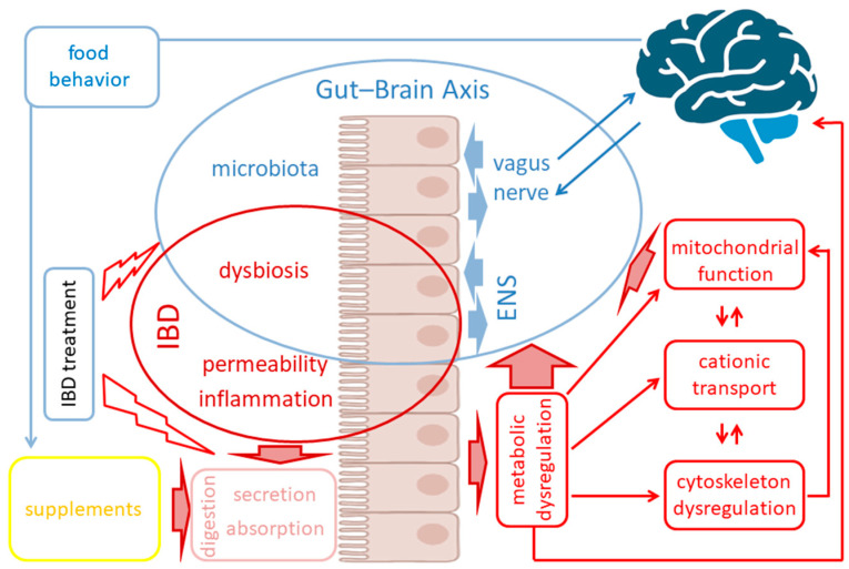
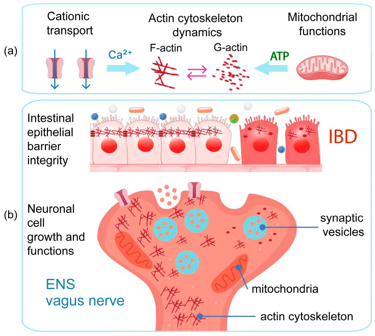
The gut-brain axis (GBA) plays a pivotal role in human health and wellness by orchestrating complex bidirectional regulation and influencing numerous critical processes within the body. Over the past decade, research has increasingly focused on the GBA in the context of inflammatory bowel disease (IBD). Beyond its well-documented effects on the GBA-enteric nervous system and vagus nerve dysregulation, and gut microbiota misbalance-IBD also leads to impairments in the metabolic and cellular functions: metabolic dysregulation, mitochondrial dysfunction, cationic transport, and cytoskeleton dysregulation. These systemic effects are currently underexplored in relation to the GBA; however, they are crucial for the nervous system cells' functioning. This review summarizes the studies on the particular mechanisms of metabolic dysregulation, mitochondrial dysfunction, cationic transport, and cytoskeleton impairments in IBD. Understanding the involvement of these processes in the GBA may help find new therapeutic targets and develop systemic approaches to improve the quality of life in IBD patients.

Keywords: IBD; cationic transport; cytoskeleton; gut–brain axis; inflammation; lipidome; metabolome; mitochondrial function.


Conflict of interest statement

The authors declare no conflicts of interest. The funders had no role in the design of the study; in the collection, analyses, or interpretation of data; in the writing of the manuscript; or in the decision to publish the results.

Figures

Figure 1
Figure 1
Schematic overview of metabolic dysregulation, mitochondrial function, cationic transport, and cytoskeletal dysregulation roles within GBA imbalance in IBD pathogenesis. Blue arrows indicate normal interactions; red arrows and broken arrows indicate negative effects of IBD-related processes and IBD treatment, accordingly.
Figure 2
Figure 2
Schematic overview of mechanisms of metabolic dysregulation impact on GBA functions in IBD. (a) Schematic presentation of IBD-induced metabolic dysregulation effects on neural cells’ functions and signaling within GBA. Red boxes indicate metabolic processes found impaired in IBD; blue boxes indicate neural cellular functions subject to metabolic dysregulation effects; red arrows indicate negative effects of IBD-related processes; (b) Schematic presentation of IBD-related lipidome dysregulation effects on cellular and organelle membranes and functions; biosignaling processes; and extracellular transport and blood–brain barrier permeability. Red boxes indicate classes of compounds whose metabolism is impaired in IBD; blue boxes indicate cellular functions subject to the corresponding lipidome dysregulation effects; red arrows indicate negative effects of lipidome dysregulation.
Figure 3
Figure 3
Mitochondrial functions’ involvement in GBA imbalance in IBD. (a) Mitochondrial health is highly crucial for neural cells’ function being subject to IBD-related pathological processes. (b) Lipid droplets and mitochondrion-associated membranes’ role in mitochondrial functions and calcium transport regulation. MAM—mitochondria-associated endoplasmic reticulum membrane, IP3R—inositol triphosphate receptor (a membrane glycoprotein complex that acts as a Ca2⁺ channel), VDAC—voltage-dependent anion channels, PLN1 and PLN5—surface proteins of lipid droplets involved in the contact of lipid droplets with mitochondria, MIGA2 is an outer mitochondrial membrane protein that directly links mitochondria to lipid droplets. (c) Mitochondria regulate actin filament dynamics that are crucial for intracellular junctions’ integrity through ATP production. Mitochondrial respiration also promotes mitochondria-associated intra-axonal translation of actin regulatory proteins involved in axonal branching. Both the mitochondria-dependent regulation of actin dynamics and intra-axonal translation are regulated by the modulation of mitochondrial respiration by metabolic signals and cytoskeleton dynamics. Blue arrows indicate the directions of influence between the interconnected processes shown in the schemes.
Figure 4
Figure 4
TRP ion channels’ functions in the gut enterocytes and neurons. Blue arrows indicate the key effects of TRP-mediated cationic transport dysregulation: calcium transport and calcium deficit significantly affects mitochondrial function’s and actin dynamics in both enterocytes and neurons; TRP ion channels perform crucial functions in enteric nervous system (ENS) neurons and regulate calcium transport and response to various signals; calcium transport affects gut epithelial integrity, and calcium deficit induces “leaky gut” syndrome; TRP ion channels and cationic transport regulate immune responses via immune cells receptors’ signaling.
Figure 5
Figure 5
Actin cytoskeleton dynamics involved both in the intestinal barrier integrity maintenance and neuronal cell functions. (a) Actin cytoskeleton dynamics and actin filament formation depends on mitochondrial ATP production and sufficient Ca2+ cellular levels. Blue arrows show influence of ATP and Ca2+ levels on the assembly and disassembly of actin filaments (F-actin) from monomeric globular actin (G-actin). (b) Actin cytoskeleton involved in intestinal barrier integrity maintenance via intercellular junctions’ stabilization. In neuronal cells, actin cytoskeleton is essential for proper vesicle formation, transmembrane receptors’ positioning and action, axonal growth, mitochondrial movement within axons, and mitophagy.

References

    1. Mayer E.A., Nance K., Chen S. The Gut-Brain Axis. Annu. Rev. Med. 2022;73:439–453. doi: 10.1146/annurev-med-042320-014032. - DOI - PubMed
    1. Barton J.R., Londregan A.K., Alexander T.D., Entezari A.A., Covarrubias M., Waldman S.A. Enteroendocrine cell regulation of the gut-brain axis. Front. Neurosci. 2023;17:1272955. doi: 10.3389/fnins.2023.1272955. - DOI - PMC - PubMed
    1. Wiley N.C., Dinan T.G., Ross R.P., Stanton C., Clarke G., Cryan J.F. The microbiota-gut-brain axis as a key regulator of neural function and the stress response: Implications for human and animal health. J. Anim. Sci. 2017;95:3225–3246. doi: 10.2527/jas.2016.1256. - DOI - PubMed
    1. Gunther C., Rothhammer V., Karow M., Neurath M., Winner B. The Gut-Brain Axis in Inflammatory Bowel Disease-Current and Future Perspectives. Int. J. Mol. Sci. 2021;22:8870. doi: 10.3390/ijms22168870. - DOI - PMC - PubMed
    1. Koloski N.A., Jones M., Kalantar J., Weltman M., Zaguirre J., Talley N.J. The brain--gut pathway in functional gastrointestinal disorders is bidirectional: A 12-year prospective population-based study. Gut. 2012;61:1284–1290. doi: 10.1136/gutjnl-2011-300474. - DOI - PubMed

LinkOut - more resources