Review
. 2024 Nov 13;25(22):12165.
doi: 10.3390/ijms252212165.

The HicAB System: Characteristics and Biological Roles of an Underappreciated Toxin-Antitoxin System

Affiliations
Review

The HicAB System: Characteristics and Biological Roles of an Underappreciated Toxin-Antitoxin System

Josefa Encina-Robles et al. Int J Mol Sci. .

Abstract

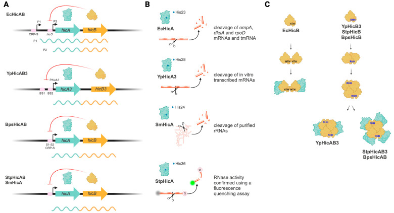
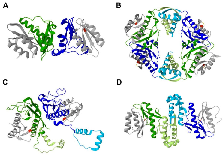
Small genetic elements known as toxin-antitoxin (TA) systems are abundant in bacterial genomes and involved in stress response, phage inhibition, mobile genetic elements maintenance and biofilm formation. Type II TA systems are the most abundant and diverse, and they are organized as bicistronic operons that code for proteins (toxin and antitoxin) able to interact through a nontoxic complex. However, HicAB is one of the type II TA systems that remains understudied. Here, we review the current knowledge of HicAB systems in different bacteria, their main characteristics and the existing evidence to associate them with some biological roles, are described. The accumulative evidence reviewed here, though modest, underscores that HicAB systems are underexplored TA systems with significant potential for future research.

Keywords: HicA; HicAB; HicB; biofilm; persistence; phage defense; plasmid maintenance; toxin-antitoxin systems; virulence.


Conflict of interest statement

The authors declare no conflicts of interest.

Figures

Figure 1
Figure 1
Genetic organization and main characteristics of characterized HicAB systems. Characteristic of E. coli HicAB (EcHicAB), Y. pestis HicAB3 (YpHicAB3), B. pseudomallei HicAB (BpsHicAB), S. meliloti HicAB (SmHicAB) and S. pneumoniae HicAB (StpHicAB) are included in the scheme. (A) Genetic organization of hicAB operons. E. coli hicAB has two promoters (P1 and P2). P1 contains a CRP-binding site (CRP-S), and it allows expression of both toxin and antitoxin genes. P2 contains the operator hicO and it is repressed by EcHicB antitoxin; when it is active, P2 allows expression of the antitoxin only. In Y. pestis hicAB, a unique upstream promoter contains two operator sequences (BS1 and BS2) to which YpHicB3 binds. B. pseudomallei hicAB possesses a putative upstream promoter with palindromic DNA binding sites (S1–2) for BpsHicB binding; S1—S2 overlap with a predicted CRP-S motif. For S. pneumonia and S. meliloti hicAB operons, HicB antitoxins were shown to bind their putative promoter, but specific binding sites are unknown. Bicistronic transcripts from BpshicAB, StphicAB and SmhicAB have not been validated. The HicA effects on HicB DNA-binding are omitted; for details, see the main text. Red curved arrows indicate inhibition. (B) HicA toxins and their described RNase activities. The experimental evidence demonstrating RNase activity for each toxin is indicated. Important catalytic residues for each HicA toxin are schematized by blue dots. (C) HicB and HicAB complex formation. HicB antitoxins have HTH (EcHicB) or RHH DNA-binding domains. EcHicB forms a single dimeric DNA-binding unit able to interact with two HicA toxins. On the contrary, RHH antitoxins (YpHicB3, StpHicB and BpsHicB) form tetramers (a dimer of dimers), containing two exposes RHH DNA-binding domains, and they interact with HicA toxins forming both heterohexameric (YpHicAB3) or heterooctameric (StpHicB and BpsHicB) complexes. Schemes are not to scale. Created in BioRender.com.
Figure 2
Figure 2
Cartoon representations of HicAB complex crystal structures. (A) YpHicAB3 (PDB: 4P78), lacking the C-terminal domain of YpHicB3, forms a heterotetramer, containing two copies of YpHicA3 and YpHicB3. (B) BpsHicAB (PDB: 6G26) is a heterooctamer, composed by four HicA and four HicB subunits. (C) StpHicAB (PDB: 5YRZ) and (D) EcHicAB (PDB: 6HPB) complexes form heterotetrameric assemblies, formed by two copies of both HicA and HicB. HicB subunits are in green and blue; HicA subunits are in grey; important catalytic residues for each HicA toxin are in red (His) or yellow (Gly22 in BpsHicAB); HicB C-terminal DNA-binding domains are highlighted in light green or light blue.

Similar articles

Cited by

References

    1. Van Melderen L. Toxin–Antitoxin Systems: Why so Many, What For? Curr. Opin. Microbiol. 2010;13:781–785. doi: 10.1016/j.mib.2010.10.006. - DOI - PubMed
    1. Pandey D.P. Toxin-Antitoxin Loci Are Highly Abundant in Free-Living but Lost from Host-Associated Prokaryotes. Nucleic Acids Res. 2005;33:966–976. doi: 10.1093/nar/gki201. - DOI - PMC - PubMed
    1. Díaz-Orejas R., Espinosa M., Yeo C.C. The Importance of the Expendable: Toxin–Antitoxin Genes in Plasmids and Chromosomes. Front. Microbiol. 2017;8:1479. doi: 10.3389/fmicb.2017.01479. - DOI - PMC - PubMed
    1. Hernández-Arriaga A.M., Chan W.T., Espinosa M., Díaz-Orejas R. Conditional Activation of Toxin-Antitoxin Systems: Postsegregational Killing and Beyond. Microbiol. Spectr. 2014;2 doi: 10.1128/microbiolspec.PLAS-0009-2013. - DOI - PubMed
    1. Hayes F., Van Melderen L. Toxins-Antitoxins: Diversity, Evolution and Function. Crit. Rev. Biochem. Mol. Biol. 2011;46:386–408. doi: 10.3109/10409238.2011.600437. - DOI - PubMed

MeSH terms

LinkOut - more resources