. 2024 Nov 13;25(22):12170.
doi: 10.3390/ijms252212170.

Nanotechnology-Driven Delivery of Caffeine Using Ultradeformable Liposomes-Coated Hollow Mesoporous Silica Nanoparticles for Enhanced Follicular Delivery and Treatment of Androgenetic Alopecia

Affiliations

Nanotechnology-Driven Delivery of Caffeine Using Ultradeformable Liposomes-Coated Hollow Mesoporous Silica Nanoparticles for Enhanced Follicular Delivery and Treatment of Androgenetic Alopecia

Nattanida Thepphankulngarm et al. Int J Mol Sci. .

Abstract

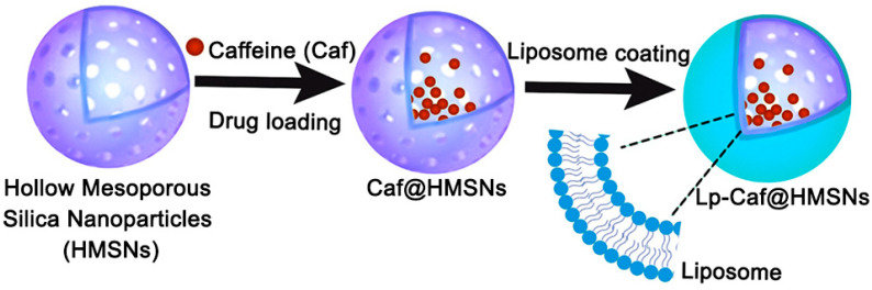
Androgenetic alopecia (AGA) is caused by the impact of dihydrotestosterone (DHT) on hair follicles, leading to progressive hair loss in men and women. In this study, we developed caffeine-loaded hollow mesoporous silica nanoparticles coated with ultradeformable liposomes (ULp-Caf@HMSNs) to enhance caffeine delivery to hair follicles. Caffeine, known to inhibit DHT formation, faces challenges in skin penetration due to its hydrophilic nature. We investigated caffeine encapsulated in liposomes, hollow mesoporous silica nanoparticles (HMSNs), and ultradeformable liposome-coated HMSNs to optimize drug delivery and release. For ultradeformable liposomes (ULs), the amount of polysorbate 20 and polysorbate 80 was varied. TEM images confirmed the mesoporous shell and hollow core structure of HMSNs, with a shell thickness of 25-35 nm and a hollow space of 80-100 nm. SEM and TEM analysis showed particle sizes ranging from 140-160 nm. Thermal stability tests showed that HMSNs coated with ULs exhibited a Td10 value of 325 °C and 70% residue ash, indicating good thermal stability. Caffeine release experiments indicated that the highest release occurred in caffeine-loaded HMSNs without a liposome coating. In contrast, systems incorporating ULp-Caf@HMSNs exhibited slower release rates, attributable to the dual encapsulation mechanism. Confocal laser scanning microscopy revealed that ULs-coated particles penetrated deeper into the skin than non-liposome particles. MTT assays confirmed the non-cytotoxicity of all HMSN concentrations to human follicle dermal papilla cells (HFDPCs). ULp-Caf@HMSNs promoted better cell viability than pure caffeine or caffeine-loaded HMSNs, highlighting enhanced biocompatibility without increased toxicity. Additionally, ULp-Caf@HMSNs effectively reduced ROS levels in DHT-damaged HFDPCs, suggesting they are promising alternatives to minoxidil for promoting hair follicle growth and reducing hair loss without increasing oxidative stress. This system shows promise for treating AGA.

Keywords: androgenetic alopecia; caffeine; confocal laser scanning microscopy (CLSM); follicular delivery; hair follicle dermal papilla cells (HFDPCs); hollow mesoporous silica nanoparticles (HMSNs); ultradeformable liposomes.


Conflict of interest statement

The authors declare no conflicts of interest.

Figures

Figure 1
Figure 1
Schematic diagram of the formation of Caf@HMSNs, Lp-Caf@HMSNs, and derivatives.
Figure 2
Figure 2
The FT-IR spectra of HMSNs and LpTW20-HMSNs.
Figure 3
Figure 3
FE-SEM images of (a,b) HMSNs and (c,d) LpTW20-HMSNs (Insets: distribution of particle size images). TEM images of (e,f) SiO2@CTAB-SiO2 Core/Shell Nanoparticles and (g,h) HMSNs after etching.
Figure 4
Figure 4
The adsorption–desorption isotherms of HMSNs (lower curve in blue is adsorption, the upper curve in red is desorption; insets show pore size distributions).
Figure 5
Figure 5
Powder XRD patterns obtained from the HCl/ethanol extraction method for as-synthesized HMSNs and LpTW20-HMSNs (lower curve in red is LpTW20-HMSNs and upper curve in blue is HMSNs).
Figure 6
Figure 6
TGA analysis of HMSNs and LpTW20-HMSNs.
Figure 7
Figure 7
The images of (a) TGA analysis and (b) FT-IR spectra of pure Caf, HMSNs, and Caf@HMSNs.
Figure 8
Figure 8
Release behavior of Caf in PBS (pH = 7.4) at 37.5 °C of LpTW20-Caf@HMSNs, LpTW80-Caf@HMSNs, LpTW2080-Caf@HMSNs, Lp-Caf@HMSNs, Caf@HMSNs, and Caf@LpTW20. Data are expressed as mean and SD (n = 3). * p < 0.05 when compared to Caf@LpTW20 and Caf@HMSNs.
Figure 9
Figure 9
Cumulative amount per area of Caf from LpTW20-Caf@HMSNs, LpTW80-Caf@HMSNs, LpTW2080-Caf@HMSNs, Lp-Caf@HMSNs, Caf@HMSNs, and Caf@LpTW20 though the porcine skin. Data are expressed as mean and SD (n = 3). * p < 0.01 when compared to Caf@LpTW20.
Figure 10
Figure 10
The CLSM images illustrate the penetration of porcine skin after treatment with (a) FITC@HMSNs for one hour, (b) LpTW20-FITC@HMSNs for one hour, (c) FITC@HMSNs for six hours, and (d) LpTW20-FITC@HMSNs for six hours. The fluorescence intensity profiles at varying skin depths for (e) Rhodamine B (568 nm) and (f) FITC (488 nm). The cross-sectional CLSM images of (g) hair follicles treated with LpTW20-FITC@HMSNs for six hours, along with the corresponding fluorescence intensity profiles within the hair follicles and the fluorescence intensity profiles across the skin layers for LpTW20-FITC@HMSNs at six hours show (h) red fluorescence from Rhodamine B labeling the particles, (i) green fluorescence from FITC, and (j) a merged image combining (h) and (i) (10× objective lens).
Figure 11
Figure 11
The effect of various Caf concentrations and formulations on HFDPCs viability. Data are expressed as mean and SD (n = 3). * p < 0.01 when compared to HMSNs.
Figure 12
Figure 12
The images of cell morphology and aggregation behavior of HFDPCs treated with Caf at concentrations of (a) 0.0125 mg mL−1 and (b) 0.0250 mg mL−1 for various time intervals (0–72 h).
Figure 13
Figure 13
The effects of MNX, 0.0125, and 0.025 mg mL−1 concentrations of pure Caf, HMSNs, Caf@HMSNs, and LpTW20-Caf@HMSNs on ROS levels in DHT-damaged HFDPCs were assessed. Fluorescence microscopy was used to capture DCF-DA images, where the intensity of green fluorescence correlates with ROS concentration.

Similar articles

Cited by

References

    1. Kaur K., Kaur J., Sharma S. Evaluation of trichoscopic findings in androgenetic alopecia and their association with disease severity. Iran. J. Dermatol. 2022;25:117–122.
    1. Khantham C., Ruksiriwanich W., Sringarm K., Prom-U-Thai C., Jamjod S., Arjin C., Muangsanguan A., Rachtanapun P., Jantanasakulwong K., Phimolsiripol Y. Effects of bioactive composition in Oryza sativa L. cv. KDML105 bran extract on gene expression related to hair cycle in human hair follicle dermal papilla cells. Agronomy. 2023;13:295. doi: 10.3390/agronomy13020295. - DOI
    1. Ring C.M., Finney R., Avram M. Lasers, lights, and compounds for hair loss in aesthetics. Clin. Dermatol. 2022;40:64–75. doi: 10.1016/j.clindermatol.2021.08.013. - DOI - PubMed
    1. Hou C., Miao Y., Wang J., Wang X., Chen C.-Y., Hu Z.-Q. Collagenase IV plays an important role in regulating hair cycle by inducing VEGF, IGF-1, and TGF-β1 expression. Drug Des. Dev. Ther. 2015;9:5373–5383. doi: 10.2147/DDDT.S89124. - DOI - PMC - PubMed
    1. Kwack M.H., Sung Y.K., Chung E.J., Im S.U., Ahn J.S., Kim M.K., Kim J.C. Dihydrotestosterone-inducible dickkopf 1 from balding dermal papilla cells causes apoptosis in follicular keratinocytes. J. Investig. Dermatol. 2008;128:262–269. doi: 10.1038/sj.jid.5700999. - DOI - PubMed