Review
. 2024 Nov 14;25(22):12226.
doi: 10.3390/ijms252212226.

Molecules That Have Rarely Been Studied in Lymphatic Endothelial Cells

Affiliations
Review

Molecules That Have Rarely Been Studied in Lymphatic Endothelial Cells

Jürgen Becker et al. Int J Mol Sci. .

Abstract

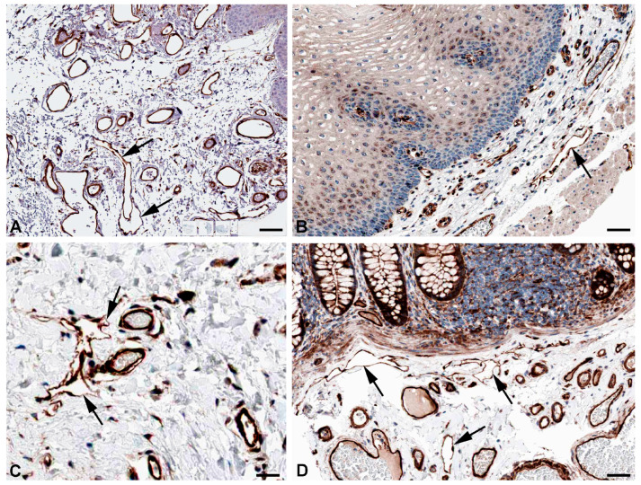
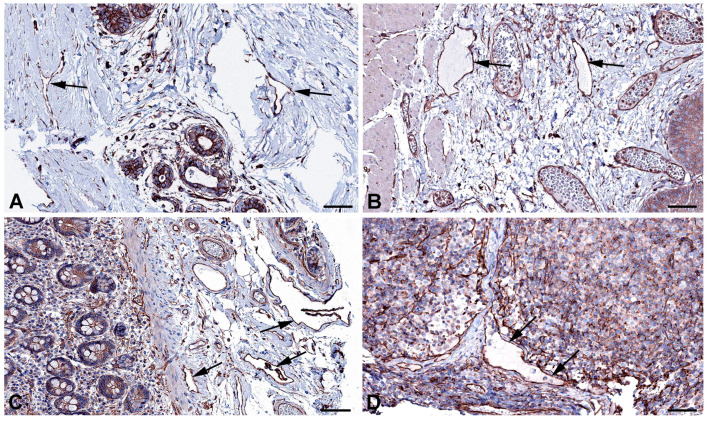
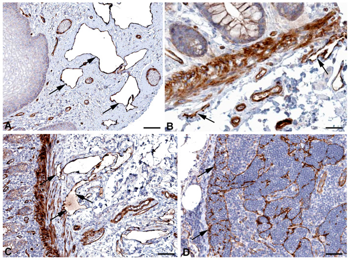
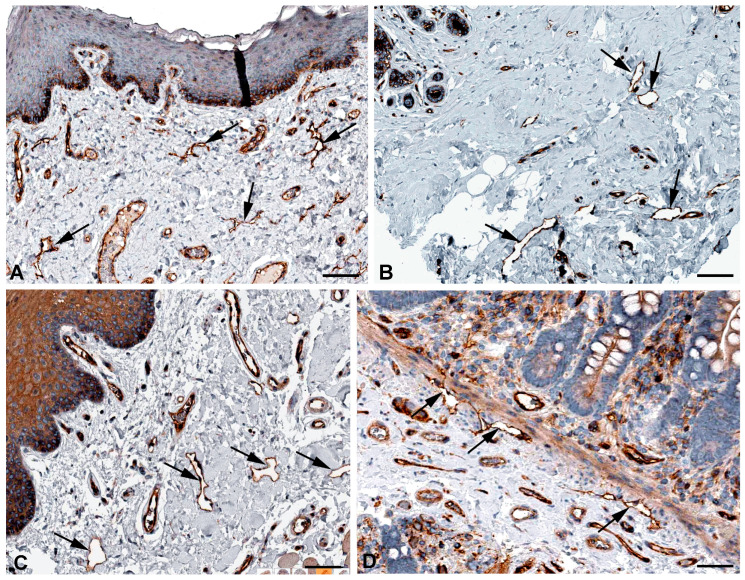
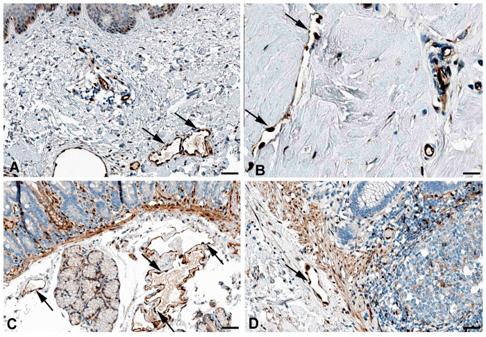
A number of standard molecules are used for the molecular and histological characterization of lymphatic endothelial cells (LECs), including lymphatic vessel endothelial hyaluronan receptor 1 (LYVE1), Podoplanin (D2-40), VEGFR3, Prospero homeobox protein 1 (PROX1), and CD31. The number of molecules whose mutations cause lymphatic malformations or primary congenital lymphedema is considerable, but the majority of these diseases have not yet been characterized at the molecular level. Therefore, there is still considerable scope for molecular and functional studies of the lymphatic vasculature. Using RNASeq, we have previously characterized lymphatic endothelial cells (LECs) under normoxic and hypoxic conditions. We used this information to compare it with immunohistochemical data. We carried out some of the immunohistology ourselves, and systematically studied the Human Protein Atlas, a cell and tissue database based in Sweden. Here we describe molecules that are expressed at RNA and protein levels in LECs, hoping to stimulate future functional studies of these molecules.

Keywords: ANKRD37; CAV1; CAV2; CD59; CNN3; DYSF; KANK3; MARCKSL1; MMRN1; NXN; SPTAN1; SPTBN1; lymphatic endothelial cell.


Conflict of interest statement

The authors declare that this study received funding from Verein zur Förderung der Lymphologie e.V., 79856 Hinterzarten, Germany. The funder was not involved in the study design, collection, analysis, interpretation of data, the writing of this article or the decision to submit it for publication.

Figures

Figure 1
Figure 1
Immunostaining of CAV1 in lymphatics (arrows) of human (A) Oral mucosa, Antibody CAB003791 (B) Rectum, Antibody CAB003791 (C) Breast, Antibody CAB003791, and (D) parietal layer of lymph node marginal sinus, Antibody HPA049326. From: The Human Protein Atlas. Bar = 60 µm in (A,D), and 40 µm in (B,C).
Figure 2
Figure 2
Immunostaining of CAV2 in lymphatics (arrows) of human (A) Esophagus, Antibody HPA044810 (B) Colon, Antibody CAB013488, (C) Oral mucosa, Antibody HPA044810, and (D) Skin, Antibody HPA044810. From: The Human Protein Atlas. Bar = 60 µm in (AD).
Figure 3
Figure 3
Immunostaining of DYSF in lymphatics (arrows) of human (A) Breast, Antibody CAB002510, (B) Duodenum, Antibody CAB002510, (C) Colon, Antibody HPA017071, (D) Skin anal, Antibody CAB002510. From: The Human Protein Atlas. Bar = 70 µm in (A,B), and 90 µm in (C,D).
Figure 4
Figure 4
Immunostaining of DYSF (red) and CD31 (green) in lymphatics of human foreskin; overview with epidermis (AC) and higher magnification of dermis (D). Blood vessels strongly express CD31; lymphatics have a punctate weak staining. Nuclei are stained blue with Dapi. In lymphatics, the red DYSF staining is dominant, but subepithelial capillaries also express DYSF. (AC) 10× objective; Bar = 100 µm, (D) 40× objective; Bar = 25 µm.
Figure 5
Figure 5
Immunostaining of SPTAN1 in lymphatics (arrows) of human (A) Oral mucosa, Antibody HPA007927, (B) Small intestine, Antibody CAB004581, (C) Breast, Antibody HPA007927, (D) Epididymis, Antibody HPA007927. From: The Human Protein Atlas. Bar = 60 µm in (A,D), and 40 µm in (B,C).
Figure 6
Figure 6
Immunostaining of SPTBN1 in lymphatics (arrows) of human (A) Esophagus, Antibody HPA013149, (B) Colon, Antibody HPA013149, (C) Breast, Antibody HPA013149. From: The Human Protein Atlas. Bar = 50 µm in (A,B), and 80 µm in (C).
Figure 7
Figure 7
Immunostaining of MMRN1 in lymphatics (arrows) of human (A) Skin, Antibody HPA035769, (B) Oral mucosa, Antibody HPA035769, (C) Urinary bladder, Antibody HPA035769. From: The Human Protein Atlas. Bar = 60 µm in (AC).
Figure 8
Figure 8
Immunostaining of MARCKSL1 in lymphatics (arrows) of human (A) Oral mucosa, Antibody HPA030528, (B) Esophagus, Antibody HPA030528, (C) Duodenum, Antibody HPA030528, (D) Cervix uteri, Antibody HPA030528. From: The Human Protein Atlas. Bar = 55 µm in (A,D), and 45 µm in (B,C).
Figure 9
Figure 9
Immunostaining of CNN3 in lymphatics (arrows) of human (A) Oral mucosa, Antibody HPA051237, (B) Esophagus, Antibody CAB009849, (C) Skin, Antibody CAB009849, (D) Colon, Antibody CAB009849. From: The Human Protein Atlas. Bar = 70 µm in (A), 40 µm in (B,D), and 30 µm in (C).
Figure 10
Figure 10
Immunostaining of ANKRD37 in lymphatics (arrows) of human (A) Breast, Antibody HPA036626, (B) Esophagus, Antibody HPA036626, (C) Colon, Antibody HPA036626, (D) Lymph node, Antibody HPA036626. From: The Human Protein Atlas. Bar = 80 µm in (AD).
Figure 11
Figure 11
Immunostaining of KANK3 in lymphatics (arrows) of human (A) Skin, (obviously edematous), Antibody HPA051153, (B) Colon, Antibody HPA051153, (C) Rectum, Antibody HPA051153, (D) Lymph node, Antibody HPA051153. From: The Human Protein Atlas. Bar = 60 µm in (A,C), 40 µm in (B), and 80 µm in (D).
Figure 12
Figure 12
Immunostaining of CD59 in lymphatics (arrows) of human (A) Skin, Antibody HPA026494, (B) Breast, Antibody HPA0264949, (C) Oral mucosa, Antibody HPA026494, (D) Colon, Antibody HPA026494. From: The Human Protein Atlas. Bar = 80 µm in (A,C), 100 µm in (B), and 50 µm in (D).
Figure 13
Figure 13
Immunostaining of NXN in lymphatics (arrows) of human (A) Skin, Antibody HPA023566, (B) Breast, Antibody HPA023566, (C) Duodenum, Antibody HPA023566, (D) Colon, Antibody HPA023566. From: The Human Protein Atlas. Bar = 60 µm in (A,C), 25 µm in (B), and 40 µm in (D).

Similar articles

References

    1. Rusznyák I., Földi M., Szabó G. Lymphologie. Physiologie und Pathologie der Lymphgefäße und des Lymphkreislaufes. Akadémiai Kiadó; Budapest, Hungary: 1969.
    1. Asellius G. De Lactibus, Sive Lacteis Venis, Quarto Vasorum Mesaraicorum Genere, Novo Invento Gasparis Asellii Cremonensis. Ex Officina Iohannis Maire 1640; Milan, Italy: 1627.
    1. Mascagni P. Vasorum Lymphaticorum Corporis Humani, Historia et Ichnographia; Senis: Ex Typographia Pazzini Carli. 1787. [(accessed on 5 November 2024)]. Available online: https://archive.org/details/ldpd_11735328_000/page/n7/mode/2up.
    1. Rudbeck O. Nova Exercitatio Anatomica, Exhibens Ductos Hepaticos Aquosos, et Vasa Glandularum Serosa (1653) Almquist and Wiksells; Uppsala, Sweden: 1930.
    1. Witte M.H., Jones K., Wilting J., Dictor M., Selg M., McHale N., Gershenwald J.E., Jackson D.G. Structure Function Relationships in the Lymphatic System and Implications for Cancer Biology. Cancer Metastasis Rev. 2006;25:159–184. doi: 10.1007/s10555-006-8496-2. - DOI - PubMed

MeSH terms

Substances

LinkOut - more resources