. 2024 Nov 14;25(22):12243.
doi: 10.3390/ijms252212243.

Relationship Between the Presence of Red Complex Species and the Distribution of Other Oral Bacteria, Including Major Periodontal Pathogens in Older Japanese Individuals

Affiliations

Relationship Between the Presence of Red Complex Species and the Distribution of Other Oral Bacteria, Including Major Periodontal Pathogens in Older Japanese Individuals

Mariko Kametani et al. Int J Mol Sci. .

Abstract

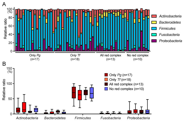
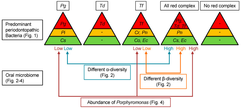
Red complex bacteria (Porphyromonas gingivalis, Treponema denticola, and Tannerella forsythia) have high virulence in periodontal disease. In the present study, we aimed to elucidate the detailed symbiotic relationships between the red complex and other oral bacteria in older Japanese individuals. Polymerase chain reaction was performed using dental plaque from 116 subjects and specific primers for ten periodontal pathogens. The detection rate of Prevotella intermedia and Capnocytophaga sputigena was significantly higher in P. gingivalis-positive subjects than in P. gingivalis-negative subjects (p < 0.05). The detection rate of Campylobacter rectus, Prevotella nigrescens, Capnocytophaga ochracea, and Eikenella corrodens was significantly higher in T. forsythia-positive subjects than in T. forsythia-negative subjects (p < 0.01). In a comprehensive analysis of oral microbiomes, three red complex species-positive subjects had significantly higher α-diversity than only P. gingivalis-positive subjects (p < 0.05) and had significantly lower β-diversity than only T. forsythia-positive subjects (p < 0.01). In the taxonomy analysis, Porphyromonas was significantly higher in three red complex species-positive subjects than in only P. gingivalis-positive and only T. forsythia-positive subjects (p < 0.01). These results suggest that each red complex species forms a unique oral microbiome and individuals positive for all red complex bacteria may harbor oral bacteria that confer a significant advantage in developing periodontal disease.

Keywords: Porphyromonas gingivalis; Tannerella forsythia; dental plaque; oral microbiome; red complex species.


Conflict of interest statement

The authors declare no conflicts of interest.

Figures

Figure 1
Figure 1
Distribution of periodontopathic bacterial species in dental plaque specimens. (A) Detection rates of each periodontopathic bacterial species. (BE) Distribution of each periodontopathic bacterial species in (B) Porphyromonas gingivalis-positive and P. gingivalis-negative subjects, (C) Treponema denticola-positive and T. denticola-negative subjects, (D) Tannerella forsythia-positive and T. forsythia-negative subjects, (E) all red complex-positive and no red complex-positive subjects. (F) Bacterial type of periodontopathic bacterial species other than red complex. * p < 0.05, ** p < 0.01, *** p < 0.001 between each group. Pg, P. gingivalis; Td, T. denticola; Tf, T. forsythia; Cr, Campylobacter rectus; Pi, Prevotella intermedia; Pn, Prevotella nigrescens; Aa, Aggregatibacter actinomycetemcomitans; Co, Capnocytophaga ochracea; Cs, Capnocytophaga sputigena; and Ec, Eikenella corrodens.
Figure 2
Figure 2
α-Diversity and β-diversity in dental plaque specimens. (AC) Analyses of α-diversity. (A) Number of operational taxonomic units (OTUs), (B) Chao1, (C) Faith’s phylogenetic diversity. Whiskers indicate maximum and minimum values, boxes indicate the interquartile range, horizontal lines within boxes indicate median values. (D,E) Analyses of β-diversity. (D) Weighted UniFrac distance. The contribution rate of each Axis (1–3) in the principal coordinate analysis is shown in parentheses. In this analysis, Axis 1 (41.8%) + Axis 2 (16.9%) + Axis 3 (9.7%) = 68.4%, which is considered to reflect approximately 70% of all the information. (E) PERMANOVA analysis of the weighted UniFrac distance. Whiskers indicate maximum and minimum values, boxes indicate the interquartile range, horizontal lines within boxes indicate median values, and circles indicate outliers. * p < 0.05 and ** p < 0.01 between each group. Pg, Porphyromonas gingivalis; Tf, Tannerella forsythia.
Figure 3
Figure 3
Taxonomic analysis at the phylum level. (A) Relative abundance of oral microbiota for each subject. (B) Relative abundance of six major phyla. Whiskers indicate maximum and minimum values, boxes indicate the interquartile range, horizontal lines within boxes indicate median values. Pg, Porphyromonas gingivalis; Tf, Tannerella forsythia.
Figure 4
Figure 4
Taxonomic analysis at the genus level. Relative abundance of (A) Actinomyces, (B) Corynebacterium, (C) Rothia, (D) Porphyromonas, (E) Prevotella, (F) Streptococcus, (G) Veillonella, (H) Neisseria, (I) Haemophilus, (J) Pseudomonas, (K) Capnocytophaga, (L) Granulicatella, (M) Fusobacterium, (N) Leptotrichia. Data expressed as the mean ± standard deviation and circles indicate each subject. Each circle represents the data of each subject. ** p < 0.01 and *** p < 0.001 between each group. Pg, Porphyromonas gingivalis; Tf, Tannerella forsythia.
Figure 5
Figure 5
Schematic representation of the relationship between red complex and other oral bacteria. Pg, Porphyromonas gingivalis; Td, Treponema denticola; Tf, Tannerella forsythia; Cr, Campylobacter rectus; Pi, Prevotella intermedia; Pn, Prevotella nigrescens; Co, Capnocytophaga ochracea; Cs, Capnocytophaga sputigena; and Ec, Eikenella corrodens.

Similar articles

Cited by

References

    1. Zhao A., Sun J., Liu Y. Understanding bacterial biofilms: From definition to treatment strategies. Front. Cell Infect. Microbiol. 2023;13:1137947. doi: 10.3389/fcimb.2023.1137947. - DOI - PMC - PubMed
    1. Wake N., Asahi Y., Noiri Y., Hayashi M., Motooka D., Nakamura S., Gotoh K., Miura J., Machi H., Iida T., et al. Temporal dynamics of bacterial microbiota in the human oral cavity determined using an in situ model of dental biofilms. NPJ Biofilms Microbiomes. 2016;2:16018. doi: 10.1038/npjbiofilms.2016.18. - DOI - PMC - PubMed
    1. Abusleme L., Hoare A., Hong B.Y., Diaz P.I. Microbial signatures of health, gingivitis, and periodontitis. Periodontol. 2000. 2021;86:57–78. doi: 10.1111/prd.12362. - DOI - PubMed
    1. Darveau R.P., Hajishengallis G., Curtis M.A. Porphyromonas gingivalis as a Potential Community Activist for Disease. J. Dent. Res. 2012;91:816–820. doi: 10.1177/0022034512453589. - DOI - PMC - PubMed
    1. Nunez-Belmar J., Morales-Olavarria M., Vicencio E., Vernal R., Cardenas J.P., Cortez C. Contribution of -Omics Technologies in the Study of Porphyromonas gingivalis during Periodontitis Pathogenesis: A Minireview. Int. J. Mol. Sci. 2022;24:620. doi: 10.3390/ijms24010620. - DOI - PMC - PubMed

MeSH terms

LinkOut - more resources