Review
. 2024 Nov 14;25(22):12252.
doi: 10.3390/ijms252212252.

Complement System and Adhesion Molecule Skirmishes in Fabry Disease: Insights into Pathogenesis and Disease Mechanisms

Affiliations
Review

Complement System and Adhesion Molecule Skirmishes in Fabry Disease: Insights into Pathogenesis and Disease Mechanisms

Albert Frank Magnusen et al. Int J Mol Sci. .

Abstract

Fabry disease is a rare X-linked lysosomal storage disorder caused by mutations in the galactosidase alpha (GLA) gene, resulting in the accumulation of globotriaosylceramide (Gb3) and its deacetylated form, globotriaosylsphingosine (Lyso-Gb3) in various tissues and fluids throughout the body. This pathological accumulation triggers a cascade of processes involving immune dysregulation and complement system activation. Elevated levels of complement 3a (C3a), C5a, and their precursor C3 are observed in the plasma, serum, and tissues of patients with Fabry disease, correlating with significant endothelial cell abnormalities and vascular dysfunction. This review elucidates how the complement system, particularly through the activation of C3a and C5a, exacerbates disease pathology. The activation of these pathways leads to the upregulation of adhesion molecules, including vascular cell adhesion molecule 1 (VCAM1), intercellular adhesion molecule 1 (ICAM1), platelet and endothelial cell adhesion molecule 1 (PECAM1), and complement receptor 3 (CR3) on leukocytes and endothelial cells. This upregulation promotes the excessive recruitment of leukocytes, which in turn exacerbates disease pathology. Targeting complement components C3a, C5a, or their respective receptors, C3aR (C3a receptor) and C5aR1 (C5a receptor 1), could potentially reduce inflammation, mitigate tissue damage, and improve clinical outcomes for individuals with Fabry disease.

Keywords: cell adhesion molecules; complement-mediated injury; endothelial dysfunction; immune cell infiltration; inflammatory cascade.


Conflict of interest statement

The authors declare no conflict of interest.

Figures

Figure 1
Figure 1
Complement pathways overview. This figure illustrates the three pathways of complement activation: the classical, lectin, and alternative pathways, each terminating in the formation of the complement 5a and membrane attack complex (MAC) and subsequent cell lysis. (a) The classical pathway is initiated when the C1 complex, composed of C1q, C1r, and C1s, binds to the Fc region of antibodies (IgM or IgG) that are attached to antigens or released from damaged cells. This binding activates the serine proteases C1r and C1s, leading to the cleavage of C4 and C2, resulting in the formation of C4b2a, a C3 convertase. (b) C4b2a cleaves C3 into C3a and C3b. C3b then binds to C4b2a to create C5 convertase (C4b2aC3b), which cleaves C5 into C5a and C5b. (c) C5b initiates the assembly of the MAC, comprising C6, C7, C8, and multiple C9 molecules, forming transmembrane channels that lead to cell lysis in targeted microorganisms or damaged host cells. (d) The lectin pathway is activated when mannose-binding lectin (MBL) or ficolins bind to carbohydrate patterns on pathogens or altered host cell surfaces. This triggers the activation of MBL-associated serine proteases (MASPs), which cleave C4 and C2, leading to the formation of C4b2a and subsequently the cleavage of C3 into C3a and C3b. The process mirrors that of the classical pathway, ultimately forming C5 convertase and the MAC. (e) The alternative pathway operates at a basal level and amplifies upon encountering pathogens or tissue injuries. It begins with the spontaneous hydrolysis of C3, forming C3 (H2O) that binds factor B, followed by cleavage by factor D to yield C3 (H2O)Bb, serving as a C3 convertase. This pathway also leads to the generation of C5 convertase and the assembly of the MAC. In the entire figure, solid, dashed, and bidirectional arrows are employed to illustrate the various components of the complement system and to show up the interconnected nature of the complement pathways, including their activation and amplification.
Figure 2
Figure 2
Mechanisms of complement activation in Fabry disease. (a) Excess accumulation of Gb3 may lead to spontaneous hydrolysis of C3 via the alternative complement activation pathway, resulting in the formation of C3 (H2O). (b). The binding of C3 (H2O) to complement factor B (CFB) initiates its cleavage by factor D (FD), producing the C3 convertase complex, C3 (H2O)Bb. This enzyme complex cleaves additional C3 molecules into C3a and C3b. (c). The generated C3b can further bind to CFB, which is then cleaved by FD to form the potent C3 convertase, C3bBb. (d) The activity of C3bBb facilitates the downstream cleavage of C3 into C3a and C3b. (e). Additionally, the interaction of C3bBb with C3b results in the formation of the C5 convertase, C3bBbC3b, leading to the cleavage of C5 into C5a within the context of Fabry disease. (f). Post-enzyme replacement therapy may trigger the production of drug-specific IgG antibodies (Drug-IgG), which bind to the therapeutic agents, agalsidase α or agalsidase β, forming drug-specific IgG immune complexes (Drug-IgG ICs). (g) The subsequent interaction of C1q with these Drug-IgG ICs activates serine proteases C1r and C1s. (h) This activation initiates the cleavage of C4 and C2, ultimately leading to the formation of the C3 convertase (C4b2a) through the classical complement pathway. (i) The resultant C3 convertase (C4b2a) facilitates the cleavage of C3 into C3a and C3b. (j) The interaction between the C3 convertase and C3b further results in the formation of the C5 convertase, which cleaves C5 into C5a, particularly in the context of Fabry disease. In the figure, dashed arrows are used to represent the components of the complement system involved in complement activation via the classical and alternative pathway in Fabry disease.
Figure 3
Figure 3
The C3a–C3aR and C5a–C5aR1 axes mediate excessive leukocyte recruitment in Fabry disease. (a,b) In Fabry disease, C3a and C5a bind to their respective receptors C3aR for C3a and C5aR1 for C5a on both leukocytes and endothelial cells. (c) Such C3a–C3aR and C5a–C5aR1 axes activation triggers a cascade of cellular responses. This interaction upregulates key adhesion molecules including P selectin, Very Late Antigen 4 (VLA 4), vascular cell adhesion molecule 1 (VCAM1), intercellular adhesion molecule 1 (ICAM1), and complement receptor 3 (CR3) on leukocytes. (d) On endothelial cells, the same receptors induce the expression of P selectin-glycoprotein ligand 1 (PSGL1), VCAM1, platelet and endothelial cell adhesion molecule 1 (PECAM1), and ICAM1. (e) The sequential activation of these adhesion molecules facilitates the progression of leukocyte recruitment. Initially, leukocytes loosely roll along the endothelial surface via P selectin–PSGL1 interactions. (fh) These rolling transitions from weaker to stronger, more stable adhesion through VLA4–VCAM1 interactions, followed by a series of firm adhesion events mediated by ICAM1-VCAM1, PECAM1, and CR3–ICAM1 interactions. (i) These interactions conclude in the transmigration of leukocytes through the endothelial barrier, exacerbating tissue damage in Fabry disease.
Figure 4
Figure 4
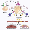
Immunological interactions and inflammatory pathways lead to vascular abnormalities in Fabry disease. (a) Circulating C3a and C5a engage their respective receptors, C3aR and C5aR, on monocytes, triggering the upregulation of the major histocompatibility complex class II (MHCII). (b) MHCII-expressing monocytes then process and present circulatory soluble intercellular adhesion molecule 1 (ICAM1) to CD4+ T cells, facilitating their activation and subsequent production of pro-inflammatory cytokines. (c) Simultaneously, MHC II-positive monocytes process and present circulatory soluble vascular cell adhesion molecule 1 (VCAM1)) to CD4+ T cells, further promoting cellular activation and enhancing pro-inflammatory cytokine production. (d) The cumulative effects of these immunological interactions and the resulting pro-inflammatory cytokines inflict damage on the glycocalyx, the protective layer of endothelial cells, leading to endothelial injury and contributing to the vascular abnormalities characteristic of Fabry disease.

Similar articles

Cited by

References

    1. Burlina A.B., Polo G., Salviati L., Duro G., Zizzo C., Dardis A., Bembi B., Cazzorla C., Rubert L., Zordan R., et al. Newborn screening for lysosomal storage disorders by tandem mass spectrometry in North East Italy. J. Inherit. Metab. Dis. 2018;41:209–219. doi: 10.1007/s10545-017-0098-3. - DOI - PubMed
    1. Gilchrist M., Casanova F., Tyrrell J.S., Cannon S., Wood A.R., Fife N., Young K., Oram R.A., Weedon M.N. Prevalence of Fabry disease-causing variants in the UK Biobank. J. Med. Genet. 2023;60:391–396. doi: 10.1136/jmg-2022-108523. - DOI - PMC - PubMed
    1. Eng C.M., Niehaus D.J., Enriquez A.L., Burgert T.S., Ludman M.D., Desnick R. Fabry disease: Twenty-three mutations including sense and antisense CpG alterations and identification of a deletional hot-spot in the α-galactosidase A gene. Hum. Mol. Genet. 1994;3:1795–1799. doi: 10.1093/hmg/3.10.1795. - DOI - PubMed
    1. Eng C.M., Resnick-Silverman L.A., Niehaus D.J., Astrin K.H., Desnick R.J. Nature and frequency of mutations in the alpha-galactosidase A gene that cause Fabry disease. Am. J. Hum. Genet. 1993;53:1186–1197. - PMC - PubMed
    1. Schäfer E., Baron K., Widmer U., Deegan P., Neumann H.P., Sunder-Plassmann G., Johansson J.-O., Whybra C., Ries M., Pastores G.M., et al. Thirty-four novel mutations of the GLA gene in 121 patients with Fabry disease. Hum. Mutat. 2005;25:412. doi: 10.1002/humu.9327. - DOI - PubMed

MeSH terms

LinkOut - more resources