. 2024 Nov 17;25(22):12326.
doi: 10.3390/ijms252212326.

Microsomal Prostaglandin E Synthase-1 Controls Colonic Prostaglandin E2 Production and Exerts a Protective Effect on Colitis Induced by Trinitrobenzene Sulfonic Acid in Mice

Affiliations

Microsomal Prostaglandin E Synthase-1 Controls Colonic Prostaglandin E2 Production and Exerts a Protective Effect on Colitis Induced by Trinitrobenzene Sulfonic Acid in Mice

Fumiaki Kojima et al. Int J Mol Sci. .

Abstract

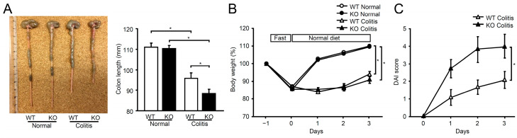
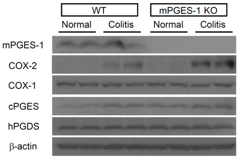
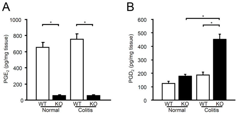
Microsomal prostaglandin E synthase-1 (mPGES-1) is an isozyme of the prostaglandin (PG) E synthase that acts downstream of cyclooxygenase and catalyzes the conversion of PGH2 to PGE2. The impact of genetic deletion of mPGES-1 on the development of 2,4,6-trinitrobenzene sulfonic acid (TNBS)-induced colitis, a well-established model of inflammatory bowel disease (IBD), was investigated in this study. After administration of TNBS, mice deficient in mPGES-1 (mPGES-1-/- mice) showed more severe colitis than did wild-type (WT) mice. Histological examination revealed that mPGES-1-/- mice had markedly exacerbated symptoms of colitis. mPGES-1 expression was detectable in the colons of WT mice at both the mRNA and protein levels. Lack of mPGES-1 resulted in marked reduction of colonic PGE2 production. Our study also showed a significant increase in colonic expression of interleukin-17A (IL-17A), as well as interferon γ (IFNγ) and tumor necrosis factor α, during colitis in mPGES-1-/- mice compared with that in WT mice. Furthermore, loss of mPGES-1 increased the populations of IL-17A-producing T-helper (Th) 17 and IFNγ-producing Th1 cells in mesenteric lymph nodes. These results suggest that mPGES-1 is the main enzyme responsible for colonic PGE2 production and deficiency of mPGES-1 facilitates the development of colitis and T-cell-mediated immunity. mPGES-1 might, therefore, impact T-cell-related immune response associated with IBD.

Keywords: Th17 and Th1 cytokines; colitis; cyclooxygenase; inflammatory bowel disease; prostaglandin E synthase; prostaglandin E2.


Conflict of interest statement

The authors declare no conflicts of interest.

Figures

Figure 1
Figure 1
Impact of mPGES-1 genetic deletion in TNBS-induced colitis. (A) On day 3 following TNBS administration, the length of each colon was assessed as an indirect indicator of inflammation (n = 11 to 13). Colon photographs are typical examples in WT and mPGES-1−/− mice. (B) Time course of body weight changes in WT and mPGES-1−/− mice before and after TNBS administration (n = 17); Mice were fasted overnight and then TNBS was administered on day 0; (C) The progression of DAI scores after the specified days of TNBS administration (n = 11 to 12). *, p < 0.05; ANOVA followed by the Bonferroni test.
Figure 2
Figure 2
Histological evaluation of TNBS-induced colitis in mPGES-1 deficiency. (A) On day 3 after TNBS administration, colon sections of mPGES-1 WT and mPGES-1−/− mice were stained with H&E. Results are typical pictures using the Swiss roll method (n = 11 to 17). Stars and arrows in the panel indicate epithelial damage and inflammatory infiltration in mPGES-1−/− mice relative to WT mice. Scale bar represents 100 µm. (B) A blinded researcher evaluated the histology scores by calculating the sum of the inflammatory infiltration score and the epithelial damage score, with a maximum possible score of 8 (n = 11 to 17). (C) On day 3 following the injection of TNBS, intestinal permeability was assessed using FITC-dextran. (n = 3 to 6). *, p < 0.05; ANOVA followed by the Bonferroni test.
Figure 3
Figure 3
Expression of mRNA for PGE2 biosynthetic enzymes in the colon following exposure to TNBS. On day 3 after TNBS administration, the expression levels of mRNA for PGES and COX isozymes in the colons were assessed using real-time RT-PCR. mRNA expression levels are presented as fold induction relative to the expression in WT mice that did not receive TNBS treatment (assigned the value “1”). *, p < 0.05; ANOVA followed by the Bonferroni test (n = 6).
Figure 4
Figure 4
Western blot analysis was conducted to examine the colonic protein expression of PGES, PGDS, and COX on day 3 following TNBS administration (n = 3).
Figure 5
Figure 5
Levels of PGE2 (A) and PGD2 (B) in the colons of mice treated or not treated with TNBS were measured using ELISA. *, p < 0.05; ANOVA followed by the Bonferroni test (n = 6 to 11).
Figure 6
Figure 6
Expression of Th17/Th1-related cytokines in the colon of mPGES-1−/− mice. On day 3 following TNBS administration, the expression of IL-17A, IFNγ, TNFα, and IL-1β mRNA in the colons of mice was examined using real-time RT-PCR. The expression levels are presented as fold changes compared to the expression in WT mice that did not receive TNBS treatment (assigned the value “1”). *, p < 0.05; ANOVA followed by the Bonferroni test (n = 6).
Figure 7
Figure 7
Population of IL-17A- and IFNγ-producing T cells in MLNs of mPGES-1−/− mice. (A) Typical FCM data showing IL-17A-producing Th17 cells and IFNγ-producing Th1 cells in MLNs of WT and mPGES-1−/− mice. On day 3 following the administration of TNBS, MLNs were isolated and assessed using FCM, as mentioned in the Materials and Methods. (B) Proportion of IL-17A+ and IFNγ+ cells in MLNs on day 3 following TNBS treatment (n = 6). *, p < 0.05 vs. WT; t test.

Similar articles

Cited by

References

    1. Friedrich M., Pohin M., Powrie F. Cytokine Networks in the Pathophysiology of Inflammatory Bowel Disease. Immunity. 2019;50:992–1006. doi: 10.1016/j.immuni.2019.03.017. - DOI - PubMed
    1. Lee S.H., Kwon J.E., Cho M.L. Immunological pathogenesis of inflammatory bowel disease. Intest. Res. 2018;16:26–42. doi: 10.5217/ir.2018.16.1.26. - DOI - PMC - PubMed
    1. Kojima F., Kapoor M., Kawai S., Crofford L.J. New insights into eicosanoid biosynthetic pathways: Implications for arthritis. Expert. Rev. Clin. Immunol. 2006;2:277–291. doi: 10.1586/1744666X.2.2.277. - DOI - PubMed
    1. Kojima F., Matnani R.G., Kawai S., Ushikubi F., Crofford L.J. Potential roles of microsomal prostaglandin E synthase-1 in rheumatoid arthritis. Inflamm. Regen. 2011;31:157–166. doi: 10.2492/inflammregen.31.157. - DOI - PMC - PubMed
    1. Jakobsson P.J., Thoren S., Morgenstern R., Samuelsson B. Identification of human prostaglandin E synthase: A microsomal, glutathione-dependent, inducible enzyme, constituting a potential novel drug target. Proc. Natl. Acad. Sci. USA. 1999;96:7220–7225. doi: 10.1073/pnas.96.13.7220. - DOI - PMC - PubMed

MeSH terms

LinkOut - more resources