

NMDA Receptors in Neurodevelopmental Disorders: Pathophysiology and Disease Models
- PMID: 39596430
- PMCID: PMC11594297
- DOI: 10.3390/ijms252212366
NMDA Receptors in Neurodevelopmental Disorders: Pathophysiology and Disease Models
Abstract
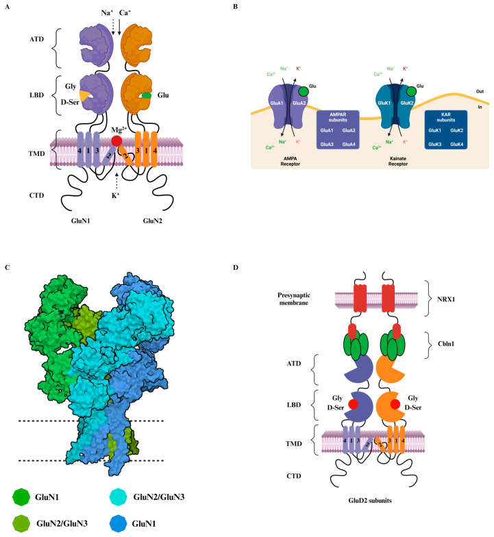
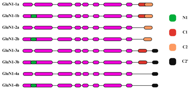
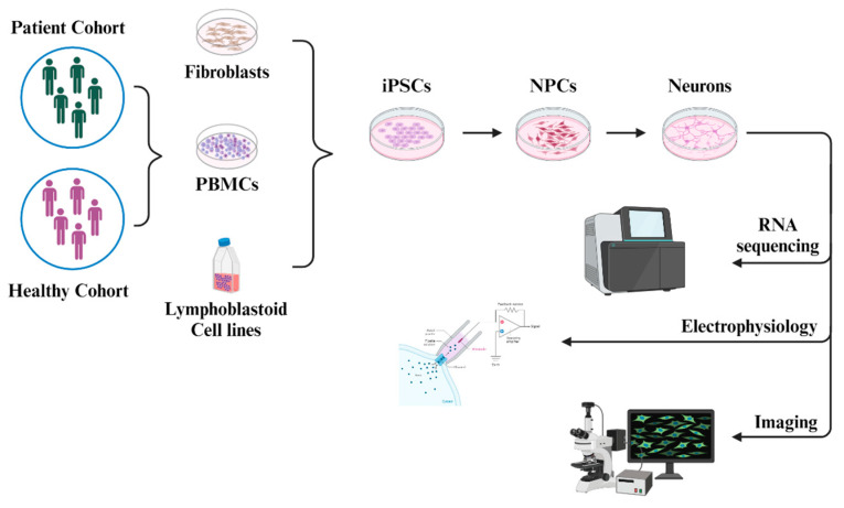
N-methyl-D-aspartate receptors (NMDARs) are critical components of the mammalian central nervous system, involved in synaptic transmission, plasticity, and neurodevelopment. This review focuses on the structural and functional characteristics of NMDARs, with a particular emphasis on the GRIN2 subunits (GluN2A-D). The diversity of GRIN2 subunits, driven by alternative splicing and genetic variants, significantly impacts receptor function, synaptic localization, and disease manifestation. The temporal and spatial expression of these subunits is essential for typical neural development, with each subunit supporting distinct phases of synaptic formation and plasticity. Disruptions in their developmental regulation are linked to neurodevelopmental disorders, underscoring the importance of understanding these dynamics in NDD pathophysiology. We explore the physiological properties and developmental regulation of these subunits, highlighting their roles in the pathophysiology of various NDDs, including ASD, epilepsy, and schizophrenia. By reviewing current knowledge and experimental models, including mouse models and human-induced pluripotent stem cells (hiPSCs), this article aims to elucidate different approaches through which the intricacies of NMDAR dysfunction in NDDs are currently being explored. The comprehensive understanding of NMDAR subunit composition and their mutations provides a foundation for developing targeted therapeutic strategies to address these complex disorders.
Keywords: ASD—autism spectrum disorder; ATD—amino-terminal domain; CNS—central nervous system; CTD—C-terminal domain; EPSCs—excitatory postsynaptic currents; LBD—ligand-binding domain; NDD—neurodevelopmental disorder; SCZ—schizophrenia; TMD—transmembrane domain; mEPSCs—miniature excitatory postsynaptic currents.
Conflict of interest statement
The authors declare no conflicts of interest.
Figures
Similar articles
-
Regulation of the NMDA receptor by its cytoplasmic domains: (How) is the tail wagging the dog?Neuropharmacology. 2021 Sep 1;195:108634. doi: 10.1016/j.neuropharm.2021.108634. Epub 2021 Jun 20. Neuropharmacology. 2021. PMID: 34097949 Free PMC article. Review.
-
Characterization of Mice Carrying a Neurodevelopmental Disease-Associated GluN2B(L825V) Variant.J Neurosci. 2024 Jul 31;44(31):e2291232024. doi: 10.1523/JNEUROSCI.2291-23.2024. J Neurosci. 2024. PMID: 38926089 Free PMC article.
-
A Frameshift Variant of GluN2A Identified in an Epilepsy Patient Results in NMDA Receptor Mistargeting.J Neurosci. 2024 Jan 24;44(4):e0557232023. doi: 10.1523/JNEUROSCI.0557-23.2023. J Neurosci. 2024. PMID: 38050135 Free PMC article.
-
Dysregulation of Neurite Outgrowth and Cell Migration in Autism and Other Neurodevelopmental Disorders.Adv Neurobiol. 2020;25:109-153. doi: 10.1007/978-3-030-45493-7_5. Adv Neurobiol. 2020. PMID: 32578146
-
De novo GRIN2A variants associated with epilepsy and autism and literature review.Epilepsy Behav. 2022 Apr;129:108604. doi: 10.1016/j.yebeh.2022.108604. Epub 2022 Feb 23. Epilepsy Behav. 2022. PMID: 35217385 Review.
Cited by
-
Exploring NMDAR pathways in ischemic stroke: implications for neurotoxic and neuroprotective mechanisms and therapeutic strategies.Naunyn Schmiedebergs Arch Pharmacol. 2025 Jun 10. doi: 10.1007/s00210-025-04357-8. Online ahead of print. Naunyn Schmiedebergs Arch Pharmacol. 2025. PMID: 40490524 Review.
-
Disclosing the Complexities of Childhood Neurodevelopmental Disorders.Children (Basel). 2024 Dec 25;12(1):16. doi: 10.3390/children12010016. Children (Basel). 2024. PMID: 39857847 Free PMC article.
References
-
- Seeburg P.H. The TiPS/TINS Lecture: The molecular biology of mammalian glutamate receptors. TiPS Rev. 1993;14:297–303. - PubMed
Publication types
MeSH terms
Substances
Grants and funding
LinkOut - more resources
Full Text Sources
Research Materials
