. 2024 Nov 18;25(22):12372.
doi: 10.3390/ijms252212372.

Tat-Beclin-1 Peptide Ameliorates Metabolic Dysfunction-Associated Steatotic Liver Disease by Enhancing Hepatic Autophagy

Affiliations

Tat-Beclin-1 Peptide Ameliorates Metabolic Dysfunction-Associated Steatotic Liver Disease by Enhancing Hepatic Autophagy

Chun-Liang Chen et al. Int J Mol Sci. .

Abstract

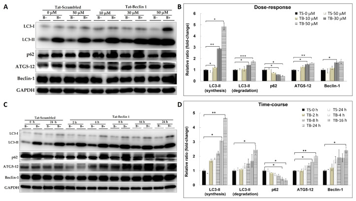
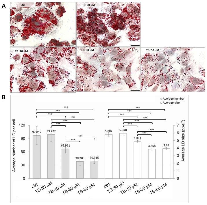
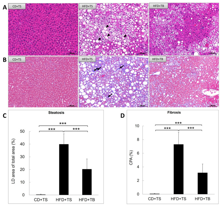
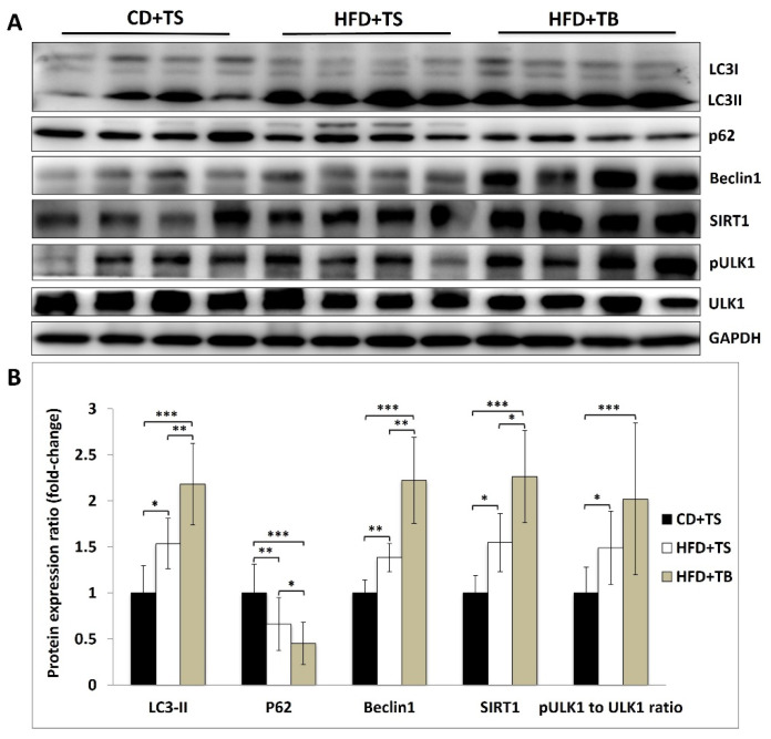
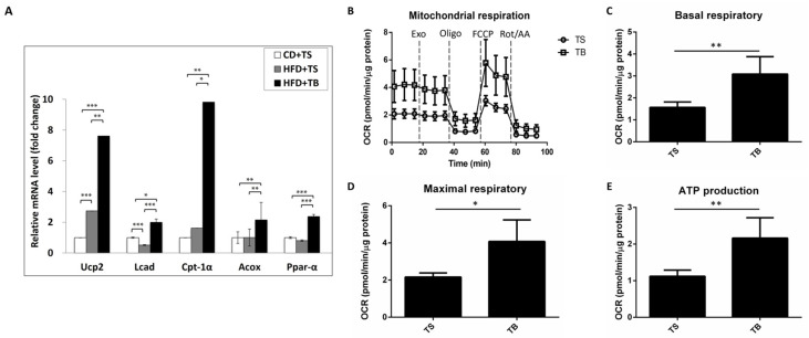
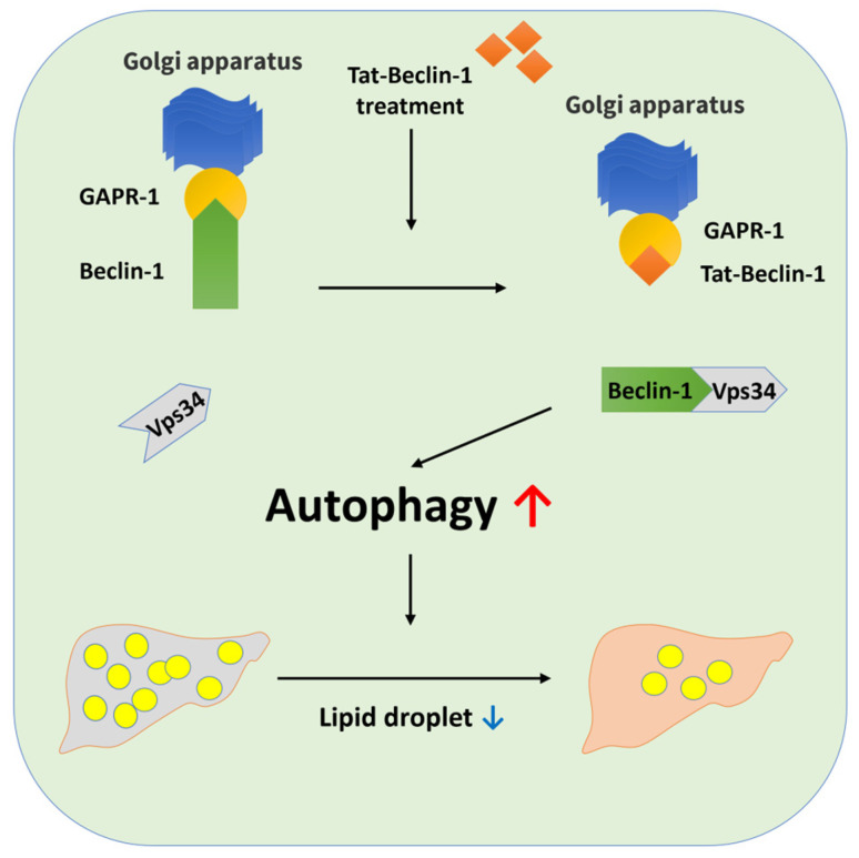
Autophagy plays a crucial role in hepatic lipid metabolism, making it a key therapeutic target for addressing metabolic dysfunction-associated steatotic liver disease (MASLD). This study evaluates the efficacy of the Tat-Beclin-1 (TB-1) peptide, a specific autophagy inducer, in mitigating MASLD. Initially, we examined the impact of the TB-1 peptide on autophagic activity and intracellular lipid metabolism in HepG2 cells treated with oleic acid, using a Tat scrambled (TS) control peptide for comparison. Subsequently, we established a MASLD mouse model by feeding a high-fat diet (HFD) for 16 weeks, followed by intraperitoneal administration of TB-1 or TS. Assessments included liver histopathology, serum biochemistry, and autophagy marker analysis. Our findings indicate that the TB-1 peptide significantly increased the LC3II/β-actin ratio in a dose- and time-dependent manner while promoting the expression of key autophagy markers Beclin-1 and ATG5-12. Furthermore, TB-1 treatment led to a marked reduction in both the size and number of lipid droplets in HepG2 cells. In vivo, HFD-fed mice exhibited increased liver weight, elevated serum alanine aminotransferase levels, and impaired oral glucose tolerance. TB-1 administration effectively mitigated these hepatic and metabolic disturbances. Histological analysis further revealed a substantial reduction in the severity of hepatic steatosis and fibrosis in TB-1-treated mice compared to TS controls. In conclusion, the TB-1 peptide shows significant potential in reducing the severity of MASLD in both HepG2 cell models and HFD-induced MASLD mouse models. Enhancing autophagy through TB-1 represents a promising therapeutic strategy for treating MASLD.

Keywords: MASLD; autophagy; fibrosis; lipid metabolism; steatosis.


Conflict of interest statement

No conflicts of interest exist for any author involved in this study.

Figures

Figure 1
Figure 1
Induction of autophagy marker expression in HepG2 cells by TB-1 peptide. HepG2 cells were treated with TB-1 peptide or TS control peptide at various concentrations (A,B) and for different durations (C,D). B+ and B− represent conditions with or without bafilomycin A1 treatment, respectively. Immunoblot analyses were conducted to quantify the levels of autophagy marker proteins. The fold change for each target protein is shown relative to the TS control sample. Statistical significance: * p < 0.05, ** p < 0.01, and *** p < 0.001. TS: Tat-Beclin-1 scrambled control peptide; TB: Tat-Beclin-1 peptide.
Figure 2
Figure 2
Reduction in intracellular lipid droplet content in HepG2 cells by TB-1 peptide. The upper panel shows Oil Red O staining of HepG2 cells treated with medium alone or TS control peptide. Treatment with TB-1 peptide at concentrations of 10, 30, and 50 μM resulted in a significant, dose-dependent decrease in lipid droplet content (red) (A). The lower panel presents the quantification of the average number and size of lipid droplets per cell (B). Lipid droplets were quantified using ImageJ software (version 1.53k), with at least 20 cells analyzed per experiment to determine the average number and size. Data are presented as the mean ± SD of three independent experiments. The scale bar represents 50 μm. Statistical significance: *** p < 0.001. TS: Tat-Beclin-1 scrambled control peptide; TB: Tat-Beclin-1 peptide.
Figure 3
Figure 3
Reduction in liver steatosis and fibrosis in the HFD-induced murine MASLD model by TB-1 peptide. Liver histology was evaluated using H and E staining to assess liver steatosis, indicated by black arrow heads (A) and Masson’s trichrome staining to evaluate liver fibrosis, indicated by black arrows (B) following treatment with the TB-1 peptide. Liver steatosis (C) and fibrosis (D) were quantified in all samples using ImageJ software (version 1.53k) and collagen proportionate area (CPA) measurement. Data are presented as mean ± SD (n = 10 per group). The scale bar represents 100 μm, with a magnification of 200×. Statistical significance: *** p < 0.001. Abbreviations: HFD, high-fat diet; CD, chow diet; H and E, hematoxylin and eosin; CPA, collagen proportionate area; LD, lipid droplet; TS, Tat scrambled control peptide; TB, Tat-Beclin-1 peptide.
Figure 4
Figure 4
Induction of autophagy in the liver of HFD-induced murine MASLD model by TB-1 peptide. (A) Protein expression levels in the liver of mice were analyzed using Western blotting. (B) The immunoblots were quantified using Image J software (version 1.53k), with results presented as mean ± SD (n = 10 per group). The blots show the effects of TB-1 peptide on the levels of autophagy-related proteins, including LC3-II, SQSTM1/p62, Beclin-1, SIRT1, and the phospho-ULK1 (Ser 317) to total ULK1 ratio. Statistical significance: * p < 0.05, ** p < 0.01, and *** p < 0.001. Abbreviations: CD, chow diet; HFD, high fat diet; TS, Tat scrambled control peptide; TB, Tat-Beclin-1 peptide; SIRT1, Sirtuin 1; ULK1, Unc-51-like kinase 1.
Figure 5
Figure 5
The effects of TB-1 peptide on hepatic fatty acid oxidation. (A) Expression levels of fatty acid oxidation-related genes in the liver of the HFD-induced murine MASLD model. (B) Oxygen consumption rate (OCR), (C) basal respiration, (D) maximal respiration, and (E) ATP production measured in oleic acid-loaded HepG2 cells following TB-1 peptide treatment. All data are presented as fold changes compared to the expression level of the CD + TS group (n = 10 per group). Statistical significance: * p < 0.05, ** p < 0.01, and *** p < 0.001. Abbreviations: CD, chow diet; HFD, high-fat diet; TS, Tat scrambled control peptide; TB, Tat-Beclin-1 peptide; Ucp2, uncoupling protein 2; Lcad, long-chain acyl-CoA dehydrogenase; Cpt-1α, carnitine palmitoyl-transferase 1α; Acox, acyl-CoA oxidase; Ppar-α, peroxisome proliferator-activated receptor α; OCR, oxygen consumption rate.
Figure 6
Figure 6
Proposed mechanism of action of TB-1 peptide in promoting autophagy, leading to reduced lipid droplet accumulation. GAPR-1: Golgi-Associated Plant Pathogenesis-Related Protein 1. Red arrow means increase and blue arrow means decrease.

Similar articles

References

    1. Fouda S., Jeeyavudeen M.S., Pappachan J.M., Jayanthi V. Pathobiology of Metabolic-Associated Fatty Liver Disease. Endocrinol. Metab. Clin. N. Am. 2023;52:405–416. doi: 10.1016/j.ecl.2023.01.001. - DOI - PubMed
    1. Dufour J.F., Anstee Q.M., Bugianesi E., Harrison S., Loomba R., Paradis V., Tilg H., Wong V.W., Zelber-Sagi S. Current therapies and new developments in NASH. Gut. 2022;71:2123–2134. doi: 10.1136/gutjnl-2021-326874. - DOI - PMC - PubMed
    1. Singh R., Kaushik S., Wang Y., Xiang Y., Novak I., Komatsu M., Tanaka K., Cuervo A.M., Czaja M.J. Autophagy regulates lipid metabolism. Nature. 2009;458:1131–1135. doi: 10.1038/nature07976. - DOI - PMC - PubMed
    1. Lin Y.C., Chang P.F., Lin H.F., Liu K., Chang M.H., Ni Y.H. Variants in the autophagy-related gene IRGM confer susceptibility to non-alcoholic fatty liver disease by modulating lipophagy. J. Hepatol. 2016;65:1209–1216. doi: 10.1016/j.jhep.2016.06.029. - DOI - PubMed
    1. Chen C.L., Lin Y.C. Autophagy Dysregulation in Metabolic Associated Fatty Liver Disease: A New Therapeutic Target. Int. J. Mol. Sci. 2022;23:10055. doi: 10.3390/ijms231710055. - DOI - PMC - PubMed

LinkOut - more resources