. 2024 Nov 18;25(22):12385.
doi: 10.3390/ijms252212385.

A Snapshot of Early Transcriptional Changes Accompanying the Pro-Neural Phenotype Switch by NGN2, ASCL1, SOX2, and MSI1 in Human Fibroblasts: An RNA-Seq Study

Affiliations

A Snapshot of Early Transcriptional Changes Accompanying the Pro-Neural Phenotype Switch by NGN2, ASCL1, SOX2, and MSI1 in Human Fibroblasts: An RNA-Seq Study

Ekaterina M Samoilova et al. Int J Mol Sci. .

Abstract

Direct pro-neural reprogramming is a conversion of differentiated somatic cells to neural cells without an intermediate pluripotency stage. It is usually achieved via ectopic expression (EE) of certain transcription factors (TFs) or other reprogramming factors (RFs). Determining the transcriptional changes (TCs) caused by particular RFs in a given cell line enables an informed approach to reprogramming initiation. Here, we characterized TCs in the human fibroblast cell line LF1 on the 5th day after EE of the single well-known pro-neural RFs NGN2, ASCL1, SOX2, and MSI1. As assessed by expression analysis of the bona fide neuronal markers nestin and beta-III tubulin, all four RFs initiated pro-neuronal phenotype conversion; analysis by RNA-seq revealed striking differences in the resulting TCs, although some pathways were overlapping. ASCL1 and SOX2 were not sufficient to induce significant pro-neural phenotype switches using our EE system. NGN2 induced TCs indicative of cell phenotype changes towards neural crest cells, neural stem cells, mature neurons, as well as radial glia, astrocytes, and oligodendrocyte precursors and their mature forms. MSI1 mainly induced a switch towards early stem-like cells, such as radial glia.

Keywords: RNA-seq; cell fate; cell reprogramming; cell therapy.


Conflict of interest statement

The authors declare no conflicts of interest.

Figures
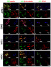
Figure 1
Figure 1
Immunocytochemical analysis of LF1 cells on day 5 after transduction. Confocal microscopy images of cells stained for nestin, β-III-tubulin, and GFAP. LF1-ASCL1 (AC), LF1-SOX2 (DF), LF1-MSI1 (GI), LF1-NGN2 (JL) samples. Green channel: GFP fluorescence confirming the integration of the lentiviral vectors. Red channel: fluorescence of the secondary antibodies (Alexa Fluor 633). The cell nuclei are stained with Hoechst (blue) in all the panels.
Figure 2
Figure 2
Differential expression analysis of RNA-seq data (A) Principal component analysis (PCA) of the log2 RNA-seq data presented as a two-dimensional scatterplot of the first two principal components. The sample groups are represented by different colors, each dot represents a biological replicate; the sample legend is provided within the plot; (B,C) Venn diagrams of the DEGs across LF1-NGN2, LF1-MSI1, and LF1-LeGoiG2 that were up- (B) and down- (C) regulated. (DG) For LF1-NGN2 and LF1-MSI1, based on each TF’s targets amongst the DEGs, the top 25 most highly affected TFs are shown ((D,F), correspondingly), as well as the estimated pathway activity ((E,G), correspondingly).
Figure 3
Figure 3
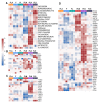
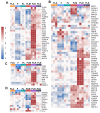
Profiles of cell subtypes in the samples based on gene expression analysis. Heatmap showing enrichment of the samples with DEGs of pro-neuronal cell populations (A), stem-like cells (B), neural crest cells (C), and radial glial cells (D). F, control fibroblasts LF1, FL, LF-LeGoiG2-Puro+, FLA, LF-LeGoiG2-Puro+-ASCL1, FLM, LF-LeGoiG2-Puro+-MSI1, FLN, LF-LeGoiG2-Puro+-NGN2, FLS, LF-LeGoiG2-Puro+-SOX2. Blue and red color scale represents the median-scaled change in gene expression or cell population enrichment (red—up, blue—down).
Figure 4
Figure 4
Profiles of cell subtypes in the samples based on gene expression analysis. Heatmap showing enrichment of the samples with DEGs of neural progenitors (A), neurons (B), presynaptic (C), and postsynaptic structures (D). F, control fibroblasts LF1, FL, LF-LeGoiG2-Puro+, FLA, LF-LeGoiG2-Puro+-ASCL1, FLM, LF-LeGoiG2-Puro+-MSI1, FLN, LF-LeGoiG2-Puro+-NGN2, FLS, LF-LeGoiG2-Puro+-SOX2. Blue and red color scale represents the median-scaled change in gene expression or cell population enrichment (red—up, blue—down).
Figure 5
Figure 5
Profiles of cell subtypes in the samples based on gene expression analysis. Heatmap showing enrichment of the samples with DEGs of astrocytes (A) and oligodendrocyte precursors (B). F, control fibroblasts LF1, FL, LF-LeGoiG2-Puro+, FLA, LF-LeGoiG2-Puro+-ASCL1, FLM, LF-LeGoiG2-Puro+-MSI1, FLN, LF-LeGoiG2-Puro+-NGN2, FLS, LF-LeGoiG2-Puro+-SOX2. Blue and red color scale represents the median-scaled change in gene expression or cell population enrichment (red—up, blue—down).

References

    1. Kwak T.H., Hali S., Kim S., Kim J., La H., Kim K.P., Hong K.H., Shin C.Y., Kim N.H., Han D.W. Robust and Reproducible Generation of Induced Neural Stem Cells from Human Somatic Cells by Defined Factors. Int. J. Stem Cells. 2020;13:80–92. doi: 10.15283/ijsc19097. - DOI - PMC - PubMed
    1. Tsunemoto R., Lee S., Szűcs A., Chubukov P., Sokolova I., Blanchard J.W., Eade K.T., Bruggemann J., Wu C., Torkamani A., et al. Diverse reprogramming codes for neuronal identity. Nature. 2018;557:375–380. doi: 10.1038/s41586-018-0103-5. - DOI - PMC - PubMed
    1. Eguchi R., Hamano M., Iwata M., Nakamura T., Oki S., Yamanishi Y. TRANSDIRE: Data-driven direct reprogramming by a pioneer factor-guided trans-omics approach. Bioinformatics. 2022;38:2839–2846. doi: 10.1093/bioinformatics/btac209. - DOI - PubMed
    1. Chanda S., Ang C.E., Davila J., Pak C., Mall M., Lee Q.Y., Ahlenius H., Jung S.W., Südhof T.C., Wernig M. Generation of induced neuronal cells by the single reprogramming factor ASCL1. Stem Cell Rep. 2014;3:282–296. doi: 10.1016/j.stemcr.2014.05.020. - DOI - PMC - PubMed
    1. Blanchard J.W., Eade K.T., Szűcs A., Lo Sardo V., Tsunemoto R.K., Williams D., Sanna P.P., Baldwin K.K. Selective conversion of fibroblasts into peripheral sensory neurons. Nat. Neurosci. 2015;18:25–35. doi: 10.1038/nn.3887. - DOI - PMC - PubMed

MeSH terms

LinkOut - more resources