. 2024 Nov 19;25(22):12390.
doi: 10.3390/ijms252212390.

Role of Balanced Involvement of the ICOS/ICOSL/Osteopontin Network in Cutaneous Wound Healing

Affiliations

Role of Balanced Involvement of the ICOS/ICOSL/Osteopontin Network in Cutaneous Wound Healing

Foteini Christaki et al. Int J Mol Sci. .

Abstract

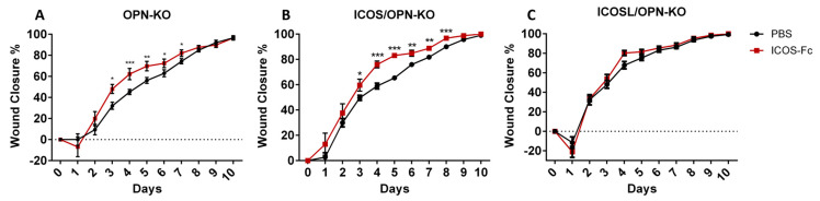
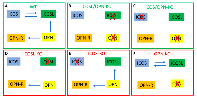
Inducible T-cell costimulator (ICOS, CD278) is a costimulatory receptor primarily expressed by activated T cells. It binds to ICOS ligand (ICOSL, CD275), which is expressed by various immune and non-immune cell types, particularly in inflamed tissues. ICOSL can also bind to osteopontin (OPN), a protein that functions both as a component of the extracellular matrix and as a soluble pro-inflammatory cytokine. Previous studies, including ours, have shown that ICOS and ICOSL play a role in skin wound healing, as mice deficient in either ICOS or ICOSL exhibit delayed healing. The aim of this study was to investigate the involvement of the ICOS/ICOSL/OPN network in skin wound healing by analyzing mice that are single knockouts for ICOS, ICOSL, or OPN, or double knockouts for ICOS/OPN or ICOSL/OPN. Our results showed that wound healing is impaired in all single knockout strains, but not in the two double knockout strains. Cellular and molecular analyses of the wound healing sites revealed that the healing defect in the single knockout strains is associated with reduced neutrophil infiltration and decreased expression of α-SMA (a marker of myofibroblasts), IL-6, TNFα, and VEGF. In contrast, the normalization of wound closure observed in the double knockout strains was primarily linked to increased vessel formation. A local treatment with recombinant ICOS-Fc improved healing in all mouse strains expressing ICOSL, but not in those lacking ICOSL, and led to a local increase in vessel formation and macrophage recruitment, predominantly of the M2 type.

Keywords: ICOS/ICOSL/OPN network; reparative macrophages; wound healing.


Conflict of interest statement

U.D. is listed among the inventors on the patent WO/2016/189428 “Ligands of B7h receptor in the treatment of osteopenia and osteoporosis” and is one of the founders of an UPO Spinoff (NOVAICOS). U.D. and C.D. listed among the inventors on the patent PCT/IB2019/050154 “Novel anti-tumor therapeutic agents”.

Figures

Figure 1
Figure 1
The wound closure rate in mice with different genotypes: osteopontin knock out (OPN-KO), Inducible T-cell costimulator knock out (ICOS-KO), ICOS ligand knock out (ICOSL-KO), ICOS/OPN-KO and ICOSL/OPN-KO. (A) The graph shows the wound closure rate of WT (n = 21) compared to OPN-KO (n = 19), ICOS-KO (n = 16), and ICOS/OPN-KO (n = 24). WT vs. ICOS/OPN-KO: * p < 0.05; ** p < 0.005; WT vs. ICOS-KO: + p < 0.05; ++ p < 0.01; +++ p < 0.0001; WT vs. OPN-KO: # p < 0.05; ## p < 0.005; ### p < 0.001. (B) The graph shows the wound closure rate of WT (n = 21) compared to OPN-KO (n = 19), ICOSL-KO (n = 18), and ICOSL/OPN-KO (n = 17). WT vs. ICOSL/OPN-KO: * p < 0.005; WT vs. ICOSL-KO: + p < 0.005; ++ p < 0.001; WT vs. OPN-KO: # p < 0.05; ## p < 0.005; ### p < 0.001. The graphs show the % of wound closure calculated using the following formula: (Wound AreaT0-Wound AreaTX)/Wound AreaT0 × 100. The statistical analysis is calculated by the Mann–Whitney test.
Figure 2
Figure 2
A comparison between WT mice and KO mice in terms of fibroblast infiltration, collagen deposition, new vessel formation (CD31+), macrophages (F4/80+), neutrophils (MPO+), and T cell (CD3+) infiltration in the wound bed (IHC). Representative images of CD31 day 3 staining are reported (20×), the arrows indicate the positive signal in correspondence of the blood vessels, n = 5 for each group. * p < 0.05; ** p < 0.005; *** p < 0.001 are calculated by the Mann–Whitney test.
Figure 3
Figure 3
A comparison between KO mice and WT mice in terms of the expression of relevant markers during the wound healing in mice treated with PBS. The graphs show the relative expression calculated by RT-PCR of different relevant markers in the wound healing: IL-6, TNFα, CD31, α-SMA, and TREM2/TREM1 ratio. (PBS n = 18; ICOS-Fc n = 18) vs. PBS WT: * p < 0.05; ** p < 0.005; *** p < 0.001 are calculated by the Mann–Whitney test.
Figure 4
Figure 4
The effect of the ICOS-Fc treatment on wound closure in OPN-KO, ICOS/OPN-KO, and ICOSL/OPN-KO mice. The graphs show the % of wound closure in (A) OPN-KO mice (n = 28–11), (B) ICOS/OPN-KO mice (n = 24–9), and (C) ICOSL/OPN-KO mice (n = 14–10) following the treatment with PBS or ICOS-Fc. The graphs show the % of wound closure calculated as previously described in Figure 2. * p < 0.05; ** p < 0.005; *** p < 0.001 are calculated by the Mann–Whitney test.
Figure 5
Figure 5
A comparison between WT mice and KO mice treated with PBS or ICOS-Fc in terms of fibroblast infiltration, collagen deposition, new vessel formation (CD31), macrophages (F4/80), neutrophils (MPO), and T cell (CD3) infiltration in the wound bed (IHC). n = 5 for each group * p < 0.05; ** p < 0.005; *** p < 0.001 are calculated by the Mann–Whitney test for the fibroblast area and CD31+ vessels and unpaired t-test for macrophages (F4/80), neutrophils (MPO), and T cell (CD3) infiltration. Representative images of CD31 day 3 staining are reported (20×), the arrows indicate the positive signal in correspondence of the blood vessels.
Figure 6
Figure 6
A comparison between KO mice in terms of the expression of relevant markers during the wound healing in mice treated with PBS and ICOS-Fc. The graphs show the relative expression calculated by RT-PCR of different relevant markers in the wound healing: IL-6, TNFα, CD31, α-SMA, and TREM/1 ratio. (PBS n = 18; ICOS-Fc n = 18). * p < 0.05; ** p < 0.005 are calculated by the Mann–Whitney test, except for the TREM2/TREM1 ratio that was calculated by the unpaired test.
Figure 7
Figure 7
Scheme of the signaling pathways triggered by the ICOS/ICOSL/OPN network in the mouse strains showing normal (green) or defective (red) wound healing. The blue arrows indicate the active signaling pathways; the red crosses indicate the defective molecules of the network; OPN-R indicate the OPN receptors other than ICOSL and include several integrins and CD44.
Figure 8
Figure 8
Scheme of the differences between the wild type mice and each knockout strain in the wound healing closure and cellular and molecular healing markers. Grey boxes: not different compared to wild type mice (WT); red boxes: decreased compared to WT mice; green boxes: increased compared to WT mice.
Figure 9
Figure 9
Scheme of the effects exerted by ICOS-Fc on the wound healing closure and cellular and molecular healing markers in wild type and knockout mice. Grey boxes: no effect induced by ICOS-Fc; red boxes: decrease induced by ICOS-Fc; green boxes: increase induced by ICOS-Fc.

Similar articles

References

    1. Singer A.J., Clark R.A. Cutaneous wound healing. N. Engl. J. Med. 1999;341:738–746. doi: 10.1056/NEJM199909023411006. - DOI - PubMed
    1. Singampalli K.L., Balaji S., Wang X., Parikh U.M., Kaul A., Gilley J., Birla R.K., Bollyky P.L., Keswani S.G. The Role of an IL-10/Hyaluronan Axis in Dermal Wound Healing. Front. Cell Dev. Biol. 2020;8:636. doi: 10.3389/fcell.2020.00636. - DOI - PMC - PubMed
    1. Aicher A., Hayden-Ledbetter M., Brady W.A., Pezzutto A., Richter G., Magaletti D., Buckwalter S., Ledbetter J.A., Clark E.A. Characterization of human inducible costimulator ligand expression and function. J. Immunol. 2000;164:4689–4696. doi: 10.4049/jimmunol.164.9.4689. - DOI - PubMed
    1. Li D., Xiong X. ICOS+ Tregs: A Functional Subset of Tregs in Immune Diseases. Front. Immunol. 2020;11:2104. doi: 10.3389/fimmu.2021.701515. Correction in Front. Immunol. 2021, 12, 701515. - DOI - PMC - PubMed
    1. Yoshinaga S.K., Whoriskey J.S., Khare S.D., Sarmiento U., Guo J., Horan T., Shih G., Zhang M., Coccia M.A., Kohno T., et al. T-cell co-stimulation through B7RP-1 and ICOS. Nature. 1999;402:827–832. doi: 10.1038/45582. - DOI - PubMed

MeSH terms

LinkOut - more resources