. 2024 Nov 19;25(22):12406.
doi: 10.3390/ijms252212406.

Gla Rich Protein (GRP) Mediates Vascular Smooth Muscle Cell (VSMC) Osteogenic Differentiation, Extracellular Vesicle (EV) Calcification Propensity, and Immunomodulatory Properties

Affiliations

Gla Rich Protein (GRP) Mediates Vascular Smooth Muscle Cell (VSMC) Osteogenic Differentiation, Extracellular Vesicle (EV) Calcification Propensity, and Immunomodulatory Properties

Carla Viegas et al. Int J Mol Sci. .

Abstract

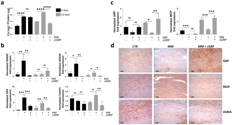
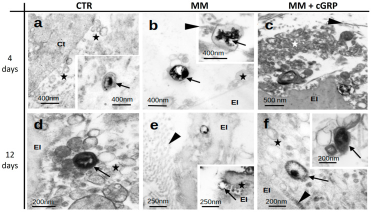
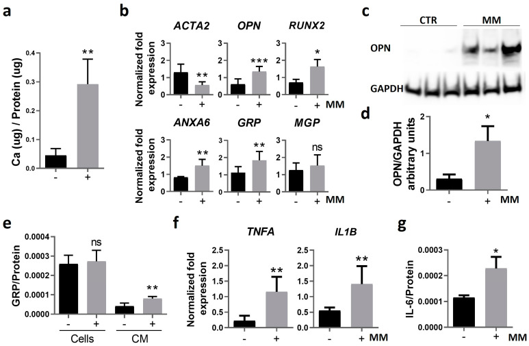
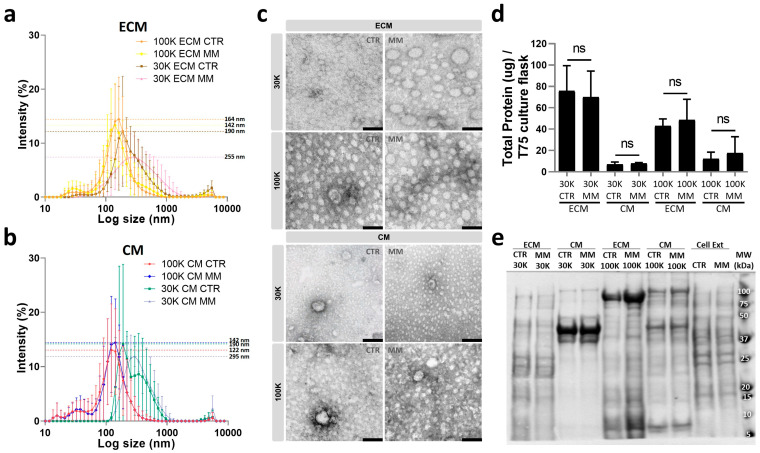
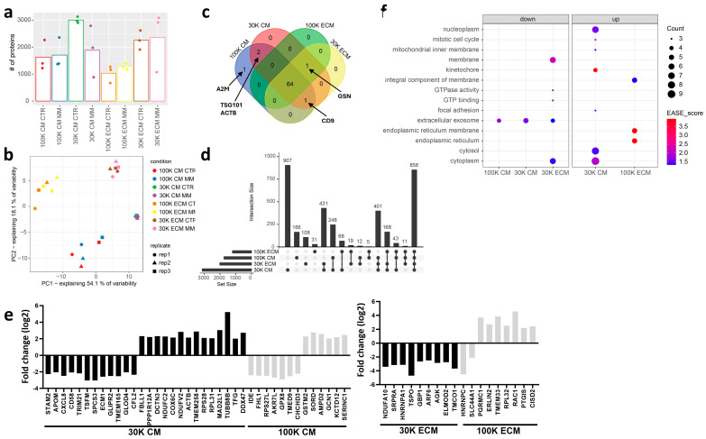
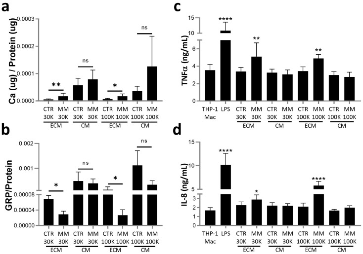
Vascular calcification (VC) is a complex process involving vascular smooth muscle cell (VSMC) osteogenic differentiation, inflammation, and extracellular vesicle (EV) calcification and communication networks. Gla rich protein (GRP) is a calcification inhibitor involved in most of these processes. However, the molecular mechanism of GRP in VC and the specific characteristics, cargo, and functionality of calcifying EVs require further elucidation. Here, we use a combination of human ex vivo aortic fragments and primary vascular smooth muscle cell (VSMC) models to obtain new information on GRP function in VC and EVs released by VSMCs. We demonstrate that GRP inhibits VSMC osteogenic differentiation through downregulation of bone-related proteins and upregulation of mineralization inhibitors, with decreased mineral crystallinity in EVs deposited into the tissue extracellular matrix (ECM). EVs isolated by ultracentrifugation at 30K and 100K from the cell media (CM) and deposited in the ECM from control (CTR) and mineralizing (MM) VSMCs were biochemically, physically, and proteomically characterized. Four different EV populations were identified with shared markers commonly present in all EVs but with unique protein cargo and specific molecular profiles. Comparative proteomics identified several regulated proteins specifically loaded into MM EV populations associated with multiple processes involved in VC. Functional analysis demonstrated that 30K and 100K ECM-MM EVs with higher calcium and lower GRP levels induced macrophage inflammation. Our findings reinforce the functional relevance of GRP in multiple VC processes and suggest that ECM EVs released under calcification stress function as a new signaling axis on the calcification-inflammation cycle.

Keywords: extracellular vesicles; gla rich protein; inflammation; vascular calcification.


Conflict of interest statement

Carla Viegas and Dina Simes are cofounders of GenoGla Diagnostics. A PCT patent application PCT/PT2009000046, is owned by the University of Algarve and the Centre of Marine Sciences (CCMAR), and the exclusive rights are licensed to GenoGla Diagnostics. The authors declare that there is no financial or non-financial conflict of interests regarding the publi-cation of this paper.

Figures

Figure 1
Figure 1
γ-carboxylated GRP (cGRP) inhibits VSMC osteogenic differentiation in ex vivo aortic fragments by downregulation of osteogenic-related genes and upregulation of mineralization inhibitors. Aortic fragments were cultured under control, mineralization (MM), and MM supplemented with cGRP media conditions for 4 (black bars) and/or 12 days (grey bars). (a) Calcium (Ca) quantification normalized to total protein levels. (b) Relative gene expression by qPCR of the osteogenic markers runt-related transcription factor 2 (RUNX2), osterix (OSX), annexin A6 (ANXA6), and the apoptotic marker caspase 3 (CASP3). (c) Relative gene expression by qPCR of the mineralization inhibitors gla rich protein (GRP) and matrix gla protein (MGP). SD was calculated from 3 independent experiments (n = 3) and ANOVA, with comparison between groups by the Dunnett test, was performed relative to the MM condition. Statistical significance was defined as p ≤ 0.05 (*), p ≤ 0.01 (**), p ≤ 0.001 (***), and p ≤ 0.0001 (****); ns, non-significant. (d) Representative immunohistochemical (IHC) experiments in consecutive tissue sections of aortic fragments cultured for 12 days in control (CTR), MM, and MM supplemented with cGRP (MM + cGRP) conditions, to detect GRP, MGP, and α-smooth muscle actin (ASMA). Gamma adjustments were performed to homogenize the panel and were applied to the entire image. Scale bar, 100 μm.
Figure 2
Figure 2
Aortic tissue calcification is associated with calcified extracellular vesicles (EVs). Representative images of transmission electron microscopy (TEM) analysis of aortic tissues of Epon-Araldite resin ultrathin sections, cultured under control (CTR) (a,d), mineralizing (MM) (b,e), and MM supplemented with cGRP (MM + cGRP) (c,f) media for 4 and 12 days. Stars indicate extracellular vesicles; arrows indicate mineralized vesicles; arrowheads indicate collagen; El, elastin; Ct, cytoplasm of smooth muscle cell.
Figure 3
Figure 3
Calcification of human primary VSMCs is characterized by osteoblastic differentiation and increased inflammation. Human primary VSMCs were cultured in control and mineralizing (MM) conditions for 14 days. (a) The mineralization rate was determined through calcium quantification normalized to protein levels. (b) Relative gene expression of VSMC osteogenic differentiation (ACTA2 encoding for ASMA, osteopontin (OPN), RUNX2, ANXA6) and mineralization inhibitor (GRP, MGP) markers by qPCR. (c) Western blot analysis for OPN detection and glyceraldehyde 3-phosphate dehydrogenase (GAPDH) as loading control. Original gels are presented in Figure S2; (d) relative OPN protein levels normalized to GAPDH using ImageJ software version V1.53f51 (arbitrary units). (e) Quantification of GRP normalized to total protein levels in cell lysates (cells) and in the cell culture media (CM) by ELISA. (f) Relative gene expression of VSMC proinflammatory cytokines (tumor necrosis factor alpha, TNFA; interleukin-1 beta, IL1B), and (g) quantification of interleukin-6 (IL-6) released to the cell media by ELISA, normalized to total protein levels. In all experiments, SD was calculated from 3 independent experiments (n = 3), and unpaired t tests were used. Statistical significance was defined as p ≤ 0.05 (*), p ≤ 0.01 (**), p ≤ 0.001 (***); ns, non-significant. In all graphs, black bars correspond to analysis at 4 days, and grey bars to 12 days.
Figure 4
Figure 4
VSMC release different extracellular vesicle (EV) populations according to isolation centrifugation force and target location. EVs released to the cell media (CM) and deposited in the extracellular matrix (ECM) by human primary VSMCs cultured in control (CTR) and mineralizing (MM) conditions were isolated by differential ultracentrifugation at 30,000× g (30K) and 100,000× g (100K). (a,b) Particle size distribution by dynamic light scattering of EVs isolated at 100K and 30K from the ECM of CTR and MM VSMC (a), and from the CM of CTR and MM VSMC (b). Data are presented with SD from 3 independent isolation experiments (n = 3); non-significant differences were found between CTR and MM EVs within each population by unpaired t tests. (c) Representative images of transmission electron microscopy (TEM) analysis of all isolated EV populations. Scale bar, 100 nm. (d) Total protein quantification of isolated EV populations normalized to T75 confluent VSMC culture flask. SD was calculated from 3 independent experiments, and unpaired t tests were used: ns, non-significant. (e) Protein profile analyzed by SDS-PAGE of all isolated EV populations and VSMC protein extracts (Cell Ext). The positions of relevant molecular mass markers (kDa) are indicated on the right side.
Figure 5
Figure 5
Proteomic characterization of VSMC EVs. (a) Protein identification rates per experimental condition (i.e., each of the eight combinations of EV subpopulation and culture condition). (b) Principal component analysis scatter plot, showing sample projections on the first two principle components. (c) Venn diagram relating the four EV populations in terms of the fraction of their protein content belonging to Vesiclepedia’s top 100 most frequently detected proteins. (d) UpSet diagram for the full protein sets detected in the four EV populations. Each column on the x-axis refers to a subset of proteins from the universe of 3774 present in a defined combination of the four EV populations (black dots). (e) Log2 fold changes of proteins whose levels are altered (adj. p-value < 0.05 and |log2 fold change| >2) in MM relative to CTR condition in each of the four sample types. (f) Bubble plot showing enriched GO terms (EASE SCORE < 0.05) in the lists of upregulated and downregulated proteins from each sample type’s MM to CTR comparison. STAM2, signal transducing adapter molecule 2; APOM, apolipoprotein M; CXCL8, interleukin-8; CD58, lymphocyte function-associated antigen 3; TRIM21, E3 ubiquitin-protein ligase TRIM21; TSFM, elongation factor Ts, mitochondrial; SPCS3, signal peptidase complex subunit 3; ECM1, extracellular matrix protein 1; GLIPR2, golgi-associated plant pathogenesis-related protein 1; TMEM165, transmembrane protein 165; GLOD4, glyoxalase domain-containing protein 4; CFL2, cofilin-2; FBLL1, rRNA/tRNA 2-O-methyltransferase fibrillarin-like protein 1; PPP1R12A, protein phosphatase 1 regulatory subunit 12A; DCTN3, dynactin subunit 3; NDUFC2, NADH dehydrogenase [ubiquinone] 1 subunit C2; COX6C, cytochrome c oxidase subunit 6C; NDUFV2, NADH dehydrogenase [ubiquinone] flavoprotein 2, mitochondrial; ACTB, actin, cytoplasmic 1; TMEM258, transmembrane protein 258; RPS28, 40S ribosomal protein S28; RPL31, 60S ribosomal protein L31; MAD2L1, mitotic spindle assembly checkpoint protein MAD2A; TUBB8B, tubulin beta-8 chain; TFG, protein TFG; DDX47, probable ATP-dependent RNA helicase DDX47; IDE, insulin-degrading enzyme; FHL1, four-and-a-half LIM domains protein 1; RPS27L, 40S ribosomal protein S27-like; AKR7L, aflatoxin B1 aldehyde reductase member 4; GPX8, glutathione peroxidase 8; TMED9, transmembrane emp24 domain-containing protein 9; CHCHD3, MICOS complex subunit MIC19; GSTM2, glutathione S-transferase Mu 2; SORD, sorbitol dehydrogenase; AMPD2, AMP deaminase 2; GCN1, eIF-2-alpha kinase activator GCN1; KCTD12, BTB/POZ domain-containing protein KCTD12; SERINC1, serine incorporator 1; NDUFA10, NADH dehydrogenase [ubiquinone] 1 alpha subcomplex subunit 10, mitochondrial; SRPRA, signal recognition particle receptor subunit alpha; HNRNPA1, heterogeneous nuclear ribonucleoprotein A1; TSPO, translocator protein; GBP1, guanylate-binding protein 1; ARF6, ADP-ribosylation factor 6; AGK, acylglycerol kinase, mitochondrial; ELMOD2, ELMO domain-containing protein 2; TMCO1, calcium load-activated calcium channel; HNRNPC, heterogeneous nuclear ribonucleoproteins C1/C2; SLC44A1, choline transporter-like protein 1; PGRMC1, membrane-associated progesterone receptor component 1; ERLIN2, erlin-2; TMEM33, transmembrane protein 33; RPL32, 60S ribosomal protein L32; RAC1, ras-related C3 botulinum toxin substrate 1; PTGIS, prostacyclin synthase; CISD2, CDGSH iron-sulfur domain-containing protein 2.
Figure 6
Figure 6
Extracellular matrix (ECM)-deposited EVs released by VSMCs under mineralization stress contain higher calcium (Ca) and decreased GRP levels and induce macrophage inflammation. (a) Calcium (Ca) content and (b) GRP quantification by ELISA, both normalized to protein levels, determined in VSMC-isolated EV populations as described in Figure 4 legend. SD was calculated from 3 independent experiments (n = 3), and unpaired t tests were used. Statistical significance was defined as p ≤ 0.05 (*), p ≤ 0.01 (**); ns, non-significant. (c,d) THP-1 differentiated macrophages (TPH-1 Mac) were treated with 5 ug/mL of VSMC EVs quantified according to total protein for each population, and the positive inflammatory control lipopolysaccharides (LPSs) (100 ng/mL), for 24 h. Levels of the proinflammatory cytokines TNFα (c) and interleukin-8 (IL-8) (d) released to the cell media were determined by ELISA. SD was calculated from 3 independent experiments (n = 3), and ANOVA, with comparison between groups by the Dunnett test, was performed relative to THP-1 Mac cells cultured in control conditions. Statistical significance was defined as p ≤ 0.05 (*), p ≤ 0.01 (**), and p ≤ 0.0001 (****).

Similar articles

Cited by

References

    1. Marreiros C., Viegas C., Simes D. Targeting a Silent Disease: Vascular Calcification in Chronic Kidney Disease. Int. J. Mol. Sci. 2022;23:16114. doi: 10.3390/ijms232416114. - DOI - PMC - PubMed
    1. Greenland P., Blaha M.J., Budoff M.J., Erbel R., Watson K.E. Coronary Calcium Score and Cardiovascular Risk. J. Am. Coll. Cardiol. 2018;72:434–447. doi: 10.1016/j.jacc.2018.05.027. - DOI - PMC - PubMed
    1. Valdivielso J.M., Rodríguez-Puyol D., Pascual J., Barrios C., Bermúdez-López M., Sánchez-Niño M.D., Pérez-Fernández M., Ortiz A. Atherosclerosis in Chronic Kidney Disease: More, Less, or Just Different? Arterioscler. Thromb. Vasc. Biol. 2019;39:1938–1966. doi: 10.1161/ATVBAHA.119.312705. - DOI - PubMed
    1. Lee S.J., Lee I.K., Jeon J.H. Vascular Calcification-New Insights Into Its Mechanism. Int. J. Mol. Sci. 2020;21:2685. doi: 10.3390/ijms21082685. - DOI - PMC - PubMed
    1. Voelkl J., Lang F., Eckardt K.U., Amann K., Kuro O.M., Pasch A., Pieske B., Alesutan I. Signaling pathways involved in vascular smooth muscle cell calcification during hyperphosphatemia. Cell. Mol. Life Sci. 2019;76:2077–2091. doi: 10.1007/s00018-019-03054-z. - DOI - PMC - PubMed

MeSH terms

Substances

LinkOut - more resources