. 2024 Nov 19;25(22):12442.
doi: 10.3390/ijms252212442.

Participation of Semaphorin Family and Plexins in the Clinical Course of Patients with Inflammatory Bowel Disease

Affiliations

Participation of Semaphorin Family and Plexins in the Clinical Course of Patients with Inflammatory Bowel Disease

Gabriela Fonseca-Camarillo et al. Int J Mol Sci. .

Abstract

Semaphorins are an immunoregulatory protein family. Plexins bind semaphorins (SEMAs) and can form receptor complexes that give them chemotactic capacity. The role and expression profile of semaphorins and plexins in inflammatory bowel disease (IBD) is currently unknown.

Aim: Characterize the semaphorins and plexins gene and protein expression in intestinal tissue from IBD patients and correlate them with the clinical phenotype.

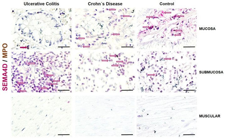
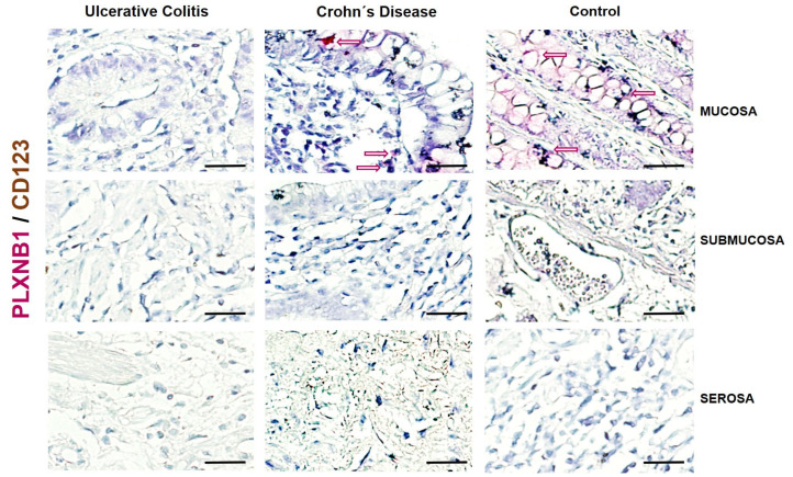
Material and methods: This comparative and cross-sectional study enrolled 54 diagnosed IBD patients and 20 controls. Gene and protein expression of semaphorins and plexins were determined by RT-PCR and IHQ for the co-localization with neutrophils (myeloperoxidase, MPO) or CD123 plasmacytoid dendritic cells in intestinal tissue from IBD patients.

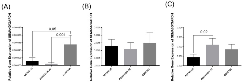
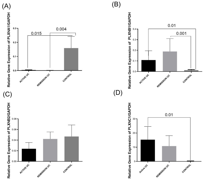
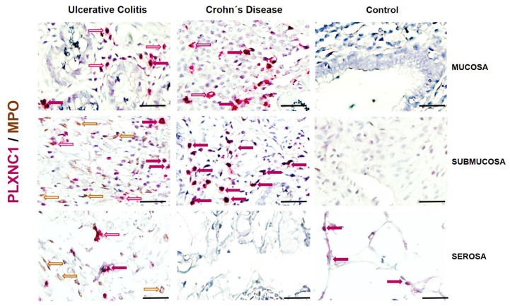
Results: Colonic mucosa from active and remission ulcerative colitis (UC) had a significantly lower SEMA4D and PLXNA1, but higher PLXNB1 gene expression than the control group. The only significant difference between active UC and remission was observed in the higher gene expression of SEMA6D in remission. It was associated with histological remission (p = 0.01, OR = 15, 95% CI: 1.39-16.1). The low expression of PLXNA1 was associated with mild intermittent activity with two relapses per year (p = 0.003, OR = 0.05, CI = 0.006-0.51). Higher SEMA4D+ positive cells were detected in the submucosa, while PLXNC1+/MPO+ in the mucosal and submucosa of active UC patients compared with controls.

Conclusions: The increased expression of the semaphorin and plexin family in IBD patients suggests their immunoregulatory function and is associated with remission and clinical phenotype in patients with UC.

Keywords: IBD; plexins; semaphorins.


Conflict of interest statement

The authors declare no conflicts of interest.

Figures

Figure 1
Figure 1
Semaphorins gene expression analysis in UC patients and controls. (A) SEMA4D gene expression, (B) SEMA5A gene expression, (C) SEMA6D gene expression. Bars show means ± S.E.M. of SEMAS transcript levels with GAPDH as the housekeeping gene. Differences among groups were assessed by the Kruskal–Wallis test. p-value < 0.05 was considered significant.
Figure 2
Figure 2
Plexins gene expression analysis in UC patients and controls. p-value < 0.05 was considered significant. (A) Plexin A1 gene expression, (B) plexin B1 gene expression, (C) plexin B2 gene expression, (D) Plexin C1 gene expression. Relative gene expression. Bars show means ± S.E.M. of plexins transcript levels with GAPDH as the housekeeping gene. Differences among groups were assessed by the Kruskal–Wallis test. p-value < 0.05 was considered significant.
Figure 3
Figure 3
Representative image of SEMA4D/MPO detection and localization in intestinal tissue from UC and CD patients and controls without intestinal inflammation. SEMA4D-positive cells were detected in pink and MPO in brown. Burgundy arrows: Double positive cells; Red outlined arrows: SEMAD4 positive cells; Brown outlined arrows: MPO positive cells. The original magnification was 600X. (Scale bar = 50 µm).
Figure 4
Figure 4
Representative image of PLXNB1/MPO detection and localization in intestinal tissue from UC and CD patients and controls without intestinal inflammation. PLXNB1-positive cells were detected in pink and MPO in brown. Burgundy arrows: Double positive cells; Red outlined arrows: PLXNB1 positive cells; Brown outlined arrows: CD123 positive cells. The original magnification was 600X. (Scale bar = 50 µm).
Figure 5
Figure 5
Representative image of PLXNC1/MPO detection and localization in intestinal tissue from UC and CD patients and controls without intestinal inflammation. PLXNC1-positive cells were detected in pink and MPO in brown. Burgundy arrows: Double positive cells; Red outlined arrows: PLXNC1 positive cells; Brown outlined arrows: MPO positive cells. The original magnification was 600X. (Scale bar = 50 µm).

Similar articles

References

    1. Podolsky D.K. Inflammatory bowel disease. N. Engl. J. Med. 2002;347:417–429. doi: 10.1056/NEJMra020831. - DOI - PubMed
    1. Cho J.H. The genetics and immunopathogenesis of inflammatory bowel disease. Nat. Rev. Immunol. 2008;8:458–466. doi: 10.1038/nri2340. - DOI - PubMed
    1. Fonseca-Camarillo G., Yamamoto-Furusho J.K. Immunoregulatory Pathways Involved in Inflammatory Bowel Disease. Inflamm. Bowel Dis. 2015;21:2188–2193. doi: 10.1097/MIB.0000000000000477. - DOI - PubMed
    1. Nishide M., Kumanogoh A. The role of semaphorins in immune responses and autoimmune rheumatic diseases. Nat. Rev. Rheumatol. 2018;14:19–31. doi: 10.1038/nrrheum.2017.201. - DOI - PubMed
    1. Kolodkin A.L., Matthes D.J., Goodman C.S. The semaphorin genes encode a family of transmembrane and secreted growth cone guidance molecules. Cell. 1993;75:1389–1399. doi: 10.1016/0092-8674(93)90625-Z. - DOI - PubMed

MeSH terms

LinkOut - more resources