. 2024 Nov 20;25(22):12443.
doi: 10.3390/ijms252212443.

Effectiveness of Local Use of Green Propolis-Loaded Lipid Nanoparticles as Adjuvant Therapy to Scaling and Root Planing in the Management of Periodontitis in Rats Treated with Zoledronate

Affiliations

Effectiveness of Local Use of Green Propolis-Loaded Lipid Nanoparticles as Adjuvant Therapy to Scaling and Root Planing in the Management of Periodontitis in Rats Treated with Zoledronate

Glauco Rodrigues Carmo Silveira et al. Int J Mol Sci. .

Abstract

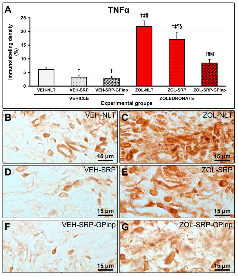
This study assessed the effectiveness of the local use of green propolis-loaded lipid nanoparticles (GPlnp) as an adjuvant therapy to scaling and root planing (SRP) to manage experimental periodontitis (EP) in ovariectomized rats treated with zoledronate. Ten weeks before the experiment, 48 female rats were ovariectomized. On day 0, a ligature was installed in the lower first molar to induce EP. From day 0 to day 42, half of the rats were treated with vehicle (VEH), while the other half were treated with 100μg/Kg of zoledronate (ZOL). On day 14, the rats were allocated into the following groups: VEH-NLT, VEH-SRP, VEH-SRP-GPlnp, ZOL-NLT, ZOL-SRP, and ZOL-SRP-GPlnp. VEH-NLT and ZOL-NLT received no local treatment. VEH-SRP and ZOL-SRP received SRP and irrigation with physiological saline solution. VEH-SRP-GPlnp and ZOL-SRP-GPlnp received SRP and irrigation with GPlnp. A single SRP session was carried out, and four irrigation sessions were conducted (on days 14, 16, 18, and 20). On day 42, all animals were euthanized. The hemimandibles were processed for histological, histometric (percentage of total bone tissue (PTBT) and non-vital bone tissue (PNVBT)) and immunohistochemical (TNFα, IL-1β, and TRAP) analysis. VEH-SRP-GPlnp showed better tissue repair, higher PTBT, and lower immunolabeling for TNFα and IL-1β compared to the groups treated with VEH. ZOL-SRP-GPlnp showed a favorable tissue repair, with lower PNVBT, less local inflammation, and lower immunolabeling for TNFα and IL-1β compared to the groups treated with ZOL. Irrigation with GPlnp proved to be effective as an adjuvant therapy to SRP in treating EP in ovariectomized rats treated with zoledronate.

Keywords: nanoparticles; periodontitis; propolis; scaling and root planing; zoledronate.


Conflict of interest statement

The authors declare no conflicts of interest.

Figures

Figure 1
Figure 1
Characterization of lipid nanoparticles containing green propolis: (A) appearance of lipid nanoparticles under a transmission electron microscope; (B) graph showing the size distribution of lipid nanoparticles (in nanometers) after 90 days of preparation; (C) average diameter of the lipid nanoparticles, zeta potential, polydispersity index, and artepelin C and baccarin encapsulation efficacies.
Figure 2
Figure 2
Bone tissue in the furcation area of the lower first molar: (A) graph showing the percentage of total bone tissue (PTBT) in the experimental groups; (BG) photomicrographs showing the histopathological characteristics in the furcation region in the (B) VEH-NLT, (C) VEH-SRP, (D) VEH-SRP-GPlnp, (E) ZOL-NLT, (F) ZOL-SRP, and (G) ZOL-SRP-GPlnp groups. In the groups treated with vehicle, it was observed that local treatments were able to reduce alveolar bone loss, especially in the VEH-SRP-GPlnp group. Note that alveolar bone loss was not severe in the groups treated with zoledronate. In the ZOL-NLT and ZOL-SRP groups, there was significant disruption of the connective tissue and extensive impairment of the bone tissue vitality. This was not observed in ZOL-SRP-GPlnp, which presented a more favorable condition. The white rectangles delimit the regions that are presented in greater magnification in Figure 3. Statistical test: Shapiro–Wilk test and variance analysis (ANOVA) followed by the Tukey post-test. Abbreviations and symbols: ab, alveolar bone; †, a statistically significant difference compared to VEH-NLT; ‡, a statistically significant difference compared to VEH-SRP; ¶, a statistically significant difference compared to VEH-SRP-GPlnp; §, a statistically significant difference compared to ZOL-NLT. Staining: Hematoxylin and Eosin. Original magnification: 50×. Scale bars: 200 μm.
Figure 3
Figure 3
Non-vital bone tissue in the furcation area of the lower first molar: (A) graph showing the percentage of non-vital bone tissue (PNVBT) in the experimental groups; (BG) photomicrographs showing the histopathological characteristics in the furcation region in the (B) VEH-NLT, (C) VEH-SRP, (D) VEH-SRP-GPlnp, (E) ZOL-NLT, (F) ZOL-SRP, and (G) ZOL-SRP-GPlnp groups. Note an intense inflammatory infiltrate in the VEH-NLT group. In contrast, the VEH-SRP and VEH-SRP-GPlnp groups presented better structured connective tissue, especially the latter. Note that in the ZOL-NLT and ZOL-SRP groups, the connective tissue was unstructured, and most of the bone tissue present was non-vital. In contrast, the ZOL-SRP-GPlnp group presented connective tissue with discrete inflammatory infiltrate, and the bone tissue presented few areas without vitality. Statistical test: Shapiro–Wilk test and variance analysis (ANOVA) followed by the Tukey post-test. Abbreviations and symbols: ab, alveolar bone; black arrows, empty lacunae; red arrows, osteocytes; †, statistically significant difference compared to VEH-NLT; ‡, statistically significant difference compared to VEH-SRP; ¶, statistically significant difference compared to VEH-SRP-GPlnp; §, statistically significant difference compared to ZOL-NLT; |, statistically significant difference compared to ZOL-SRP. Staining: Hematoxylin and Eosin. Original magnification: 100×. Scale bars: 100 μm.
Figure 4
Figure 4
Immunolabeling for TNFα in the connective tissue of the furcation region: (A) Graphs showing the immunolabeling pattern for TNFα in the experimental groups. Statistical test: Shapiro–Wilk test and variance analysis (ANOVA) followed by the Tukey post-test. (BG) Photomicrographs showing the immunolabeling patterns for TNFα in the mandibular first lower molar in the (B) VEH-NLT, (C) ZOL-NLT, (D) VEH-SRP, (E) ZOL-SRP, (F) VEH-SRP-GPlnp, and (G) ZOL-SRP-GPlnp groups. Note a higher density of immunolabeling for TNFα in the ZOL-NLT and ZOL-SRP groups. In the ZOL-SRP-GPlnp group, the local use of GPlnp as an adjuvant therapy to SRP resulted in a lower density of immunolabeling for TNFα. Symbols: †, statistically significant difference compared to VEH-NLT; ‡, statistically significant difference compared to VEH-SRP; ¶, a statistically significant difference compared to VEH-SRP-GPlnp; §, a statistically significant difference compared to ZOL-NLT; |, a statistically significant difference compared to ZOL-SRP. Original magnification: 1000×. Scale bars: 15 μm.
Figure 5
Figure 5
Immunolabeling for IL-1β in the connective tissue of the furcation region: (A) Graphs showing the immunolabeling pattern for IL-1β in the experimental groups. Statistical test: Shapiro–Wilk test and variance analysis (ANOVA) followed by the Tukey post-test. (BG) Photomicrographs showing the immunolabeling patterns for IL-1β in the mandibular first lower molar in the (B) VEH-NLT, (C) ZOL-NLT, (D) VEH-SRP, (E) ZOL-SRP, (F) VEH-SRP-GPlnp, and (G) ZOL-SRP-GPlnp groups. Note the higher density of immunolabeling for IL-1β in the ZOL-NLT and ZOL-SRP groups. In the ZOL-SRP-GPlnp group, the local use of GPlnp as an adjuvant therapy to SRP resulted in a lower density of immunolabeling for IL-1β. Symbols: †, statistically significant difference compared to VEH-NLT; ‡, statistically significant difference compared to VEH-SRP; ¶, a statistically significant difference compared to VEH-SRP-GPlnp; §, a statistically significant difference compared to ZOL-NLT; |, a statistically significant difference compared to ZOL-SRP. Original magnification: 1000×. Scale bars: 15 μm.
Figure 6
Figure 6
Immunolabeling for TRAP in the furcation region: (A) Graph showing the number of TRAP-positive cells in the furcation area of the experimental groups. Statistical test: Shapiro–Wilk test and variance analysis (ANOVA) followed by the Tukey post-test. (BG) Photomicrographs showing the immunolabeling patterns for TRAP in the mandibular left molars in the (B) VEH-NLT, (C) VEH-SRP, (D) VEH-SRP-GPlnp, (E) ZOL-NLT, (F) ZOL-SRP, and (G) ZOL-SRP-GPlnp groups. Note the higher number of TRAP-positive cells in the VEH-NLT group. In the groups treated with zoledronate, observe that the TRAP-positive cells were larger than in the groups treated with vehicle, circumferential, hypernucleated, and distant from the bone matrix. Abbreviations and symbols: ab, alveolar bone; black arrows, TRAP-positive cells (osteoclasts); †, a statistically significant difference compared to VEH-NLT. Counterstain: Harris Hematoxylin. Original magnification: 1000×. Scale bars: 20 μm.
Figure 7
Figure 7
Study design: (A) scheme depicting the procedures performed over time in the experimental groups; (B) clinical aspect two weeks after ligature placement; (C) clinical aspect after the removal of the ligature; (D) local use of green propolis-loaded lipid nanoparticles.

Similar articles

Cited by

References

    1. Ruggiero S.L., Dodson T.B., Aghaloo T., Carlson E.R., Ward B.B., Kademani D. American Association of Oral and Maxillofacial Surgeons’ Position Paper on Medication-Related Osteonecrosis of the Jaws-2022 Update. J. Oral. Maxillofac. Surg. 2022;80:920–943. doi: 10.1016/j.joms.2022.02.008. - DOI - PubMed
    1. Tetradis S., Allen M.R., Ruggiero S.L. Pathophysiology of Medication-Related Osteonecrosis of the Jaw-A Minireview. JBMR Plus. 2023;7:e10785. doi: 10.1002/jbm4.10785. - DOI - PMC - PubMed
    1. Murphy J., Mannion C.J. Medication-related osteonecrosis of the jaws and quality of life: Review and structured analysis. Br. J. Oral. Maxillofac. Surg. 2020;58:619–624. doi: 10.1016/j.bjoms.2020.03.010. - DOI - PubMed
    1. Halpern L.R., Adams D.R. Treatment of medication-related osteonecrosis of the jaw: Controversies in causality and therapy. Dent. Clin. N. Am. 2024;68:67–85. doi: 10.1016/j.cden.2023.07.005. - DOI - PubMed
    1. Beth-Tasdogan N.H., Mayer B., Hussein H., Zolk O., Peter J.U. Interventions for managing medication-related osteonecrosis of the jaw. Cochrane Database Syst. Rev. 2022;7:CD012432. doi: 10.1002/14651858.CD012432.pub3. - DOI - PMC - PubMed

LinkOut - more resources