. 2024 Nov 20;25(22):12471.
doi: 10.3390/ijms252212471.

Astrocyte Dysfunction Reflected in Ischemia-Induced Astrocyte-Derived Extracellular Vesicles: A Pilot Study on Acute Ischemic Stroke Patients

Affiliations

Astrocyte Dysfunction Reflected in Ischemia-Induced Astrocyte-Derived Extracellular Vesicles: A Pilot Study on Acute Ischemic Stroke Patients

Timea Forró et al. Int J Mol Sci. .

Abstract

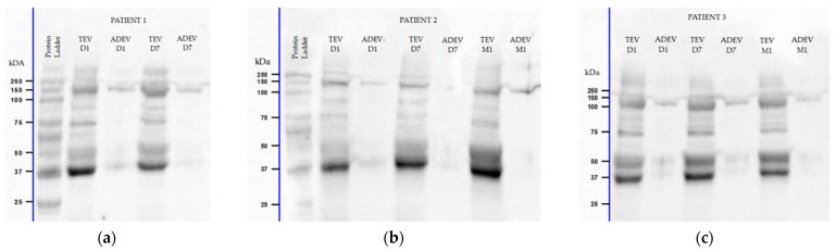
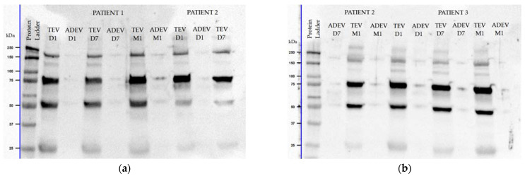
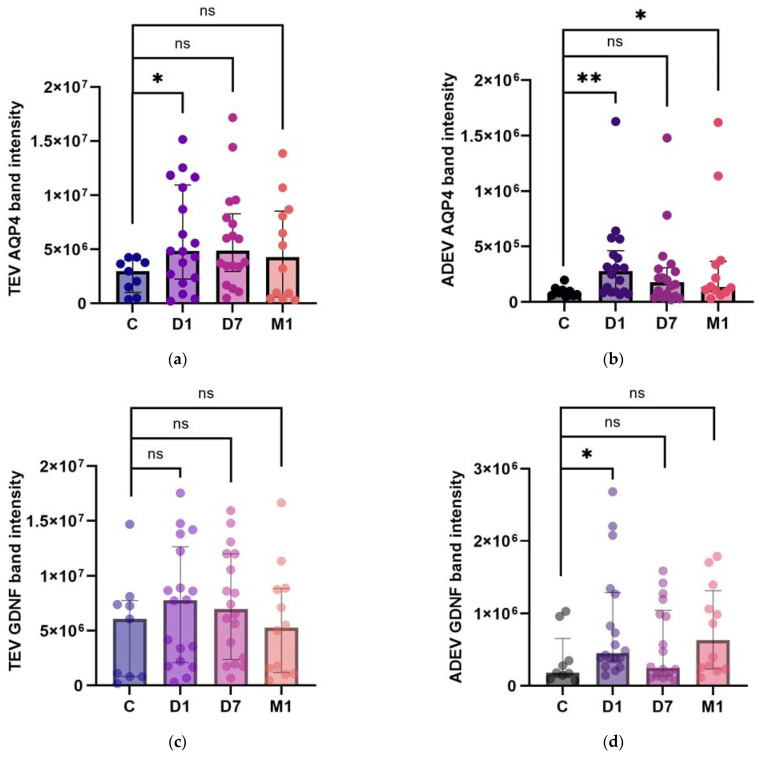
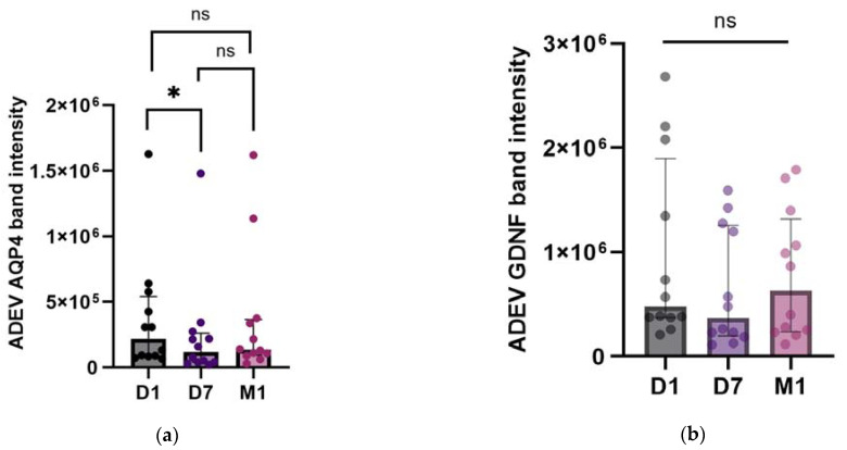
Extracellular vesicles (EVs) secreted by astrocytes (ADEVs) mediate numerous biological processes, providing insights into damage, repair, and protection following ischemic stroke (IS). This pilot study aimed to broaden the current knowledge on the astrocyte response to ischemia by dynamically assessing the aquaporin-4 (AQP4) and glial cell line-derived neurotrophic factor (GDNF) as cargo proteins of these vesicles in eighteen acute IS patients and nine controls. EV proteins were detected by Western blotting and followed 24 h (D1), 7 days (D7), and one month (M1) after symptoms onset. The post-ischemic ADEV AQP4 and GDNF levels were higher at D1 compared to the control group (p = 0.006 and p = 0.023). Significant differences were observed in ADEV AQP4 during the three evaluated time points (n = 12, p = 0.013) and between D1 and D7 (z = 2.858, p = 0.012), but not in EV GDNF. There was a positive relationship between the severity of stroke at D1 according to the National Institutes of Health Stroke Scale, and ADEV AQP4 at D1 (r = 0.50, p = 0.031), as well as ADEV GDNF at D1 and D7 (r = 0.49, p = 0.035 and r = 0.53, p = 0.021, respectively). The release of EVs with distinct protein profiles can be an attractive platform for the development of biomarkers in IS.

Keywords: Western blotting; acute ischemic stroke; aquaporin-4; astrocyte-derived extracellular vesicles; glial cell line-derived neurotrophic factor.


Conflict of interest statement

The authors declare no conflicts of interest.

Figures

Figure 1
Figure 1
Representative Western blots for aquaporin-4 (AQP4) showing multiple bands in extracellular vesicle (EV) aliquots of an AIS patient with two key moments that missed one-month follow-up (a) and two patients with three key moments (b,c): one major band at ≈37 kilodaltons (kDa), additional bands at 50 and 75 kDa, suggesting AQP4 dimers or glycosylation, and between 100–150 kDa as possible tetramers. Approximate molecular weight (MW) markers in kDa are labeled adjacent on the left. Abbreviations: TEV—total extracellular vesicles; ADEV—astrocyte-derived extracellular vesicles; D1—24 h; D7—7 days; M1—one month after stroke onset.
Figure 2
Figure 2
Representative Western blots for glial cell line-derived neurotrophic factor (GDNF) of three AIS patients with three key moments (a,b). Data for patient 2 is presented on separate blots: D1 and D7 TEV (a), D7 ADEV and M1 (b). The blots display multiple bands in EV aliquots: at ≈25 kDa as a monomer, at ≈50 kDa as a dimer, and, additionally, near 75 and 150 kDa MW as a combination of a monomer and dimer. Approximate MW markers in kDa are labeled adjacent on the left.
Figure 3
Figure 3
AQP4 (a,b) and GDNF (c,d) band intensities in TEVs (a,c) and ADEVs (b,d) of AIS patients and control participants (C). Data are represented as individual value boxplots with median and interquartile range (IQR, Mann–Whitney U test; * p  <  0.05; ** p  <  0.01; ns—not significant).
Figure 4
Figure 4
AQP4 (a) and GDNF (b) band intensities in ADEVs during the patients’ follow-up: D1, D7, and M1. Data are represented as individual value boxplots with median and IQR (Friedman’s ANOVA, followed by Dunn’s post hoc only for AQP4 (a), as for GDNF, there were no statistically significant differences (b); * p  <  0.05; ns—not significant).
Figure 5
Figure 5
Bead flow separation data for the anti-tetraspanin antibodies coupled with Exo-FITC staining showing beads with no captured EVs (a) and beads with captured EVs = tetraspanin-positive (CD9, CD63, and CD81) EVs population (b).
Figure 6
Figure 6
Bead flow separation data for the anti-GLAST antibody coupled with Exo-FITC staining showing beads with no captured EVs (a) and beads with captured EVs = GLAST-positive EVs population (b).
Figure 7
Figure 7
Transmission (TE, (a,b)) and scanning electron micrographs (SE, (ce)) of the isolated EVs shown at two different magnifications of 50,000× (a,c) and 150,000× (b,d,e). Multiple vesicles with typical EV morphology can be seen in each image.

Similar articles

Cited by

References

    1. Feigin V.L., Brainin M., Norrving B., Martins S., Sacco R.L., Hacke W., Fisher M., Pandian J., Lindsay P. World Stroke Organization (WSO): Global Stroke Fact Sheet 2022. Int. J. Stroke. 2022;17:18–29. doi: 10.1177/17474930211065917. - DOI - PubMed
    1. Feigin V.L., Stark B.A., Johnson C.O., Roth G.A., Bisignano C., Abady G.G., Abbasifard M., Abbasi-Kangevari M., Abd-Allah F., Abedi V., et al. Global, Regional, and National Burden of Stroke and Its Risk Factors, 1990–2019: A Systematic Analysis for the Global Burden of Disease Study 2019. Lancet Neurol. 2021;20:795–820. doi: 10.1016/S1474-4422(21)00252-0. - DOI - PMC - PubMed
    1. Wechsler L.R., Adeoye O., Alemseged F., Bahr-Hosseini M., Deljkich E., Favilla C., Fisher M., Grotta J., Hill M.D., Kamel H., et al. Most Promising Approaches to Improve Stroke Outcomes: The Stroke Treatment Academic Industry Roundtable XII Workshop. Stroke. 2023;54:3202–3213. doi: 10.1161/STROKEAHA.123.044279. - DOI - PubMed
    1. Kakkar P., Kakkar T., Patankar T., Saha S. Current Approaches and Advances in the Imaging of Stroke. Dis. Model. Mech. 2021;14:dmm048785. doi: 10.1242/dmm.048785. - DOI - PMC - PubMed
    1. Oberheim N.A., Goldman S.A., Nedergaard M. Heterogeneity of Astrocytic Form and Function. Methods Mol. Biol. 2012;814:23. doi: 10.1007/978-1-61779-452-0_3. - DOI - PMC - PubMed

LinkOut - more resources