. 2024 Nov 14;12(11):2311.
doi: 10.3390/microorganisms12112311.

Exiguolysin, a Novel Thermolysin (M4) Peptidase from Exiguobacterium oxidotolerans

Affiliations

Exiguolysin, a Novel Thermolysin (M4) Peptidase from Exiguobacterium oxidotolerans

Brendan F Gilmore et al. Microorganisms. .

Abstract

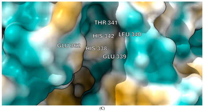
This study details a comprehensive biochemical and structural characterization of exiguolysin, a novel thermolysin-like, caseinolytic peptidase secreted by a marine isolate of Exiguobacterium oxidotolerans strain BW26. Exiguolysin demonstrated optimal proteolytic activity at 37 °C and pH 3, retaining 85% activity at 50 °C, highlighting its potential stability under broad reaction conditions. SDS-PAGE and LC-MS analysis identified the enzyme as a 32 kDa M4-family metalloprotease. Exiguolysin activity was inhibited by 1,10-phenanthroline, confirming its dependence on metal ions for activity. Zymographic analysis and substrate specificity assays revealed selective hydrolysis of matrix metalloproteinase (MMP) substrates but no activity against elastase substrates. Analysis of the predicted gene sequence and structural predictions using AlphaFold identified the presence and position of HEXXH and Glu-Xaa-Xaa-Xaa-Asp motifs, crucial for zinc binding and catalytic activity, characteristic of 'Glu-zincins' and members of the M4 peptidase family. High-throughput screening of a 20 × 20 N-alpha mercaptoamide dipeptide inhibitor library against exiguolysin identified SH-CH2-CO-Met-Tyr-NH2 as the most potent inhibitor, with a Ki of 1.95 μM. Notably, exiguolysin selectively inhibited thrombin-induced PAR-1 activation in PC-3 cells, potentially indicating a potential mechanism of virulence in modulating PAR-1 signalling during infection by disarming PARs. This is the first detailed characterization of a peptidase of the M4 (thermolysin) family in the genus Exiguobacterium which may have industrial application potential and relevance as a putative virulence factor.

Keywords: Exiguobacterium oxidotolerans; metalloprotease; protease-activated receptors; thermolysin.


Conflict of interest statement

The authors declare no conflicts of interest.

Figures

Figure 1
Figure 1
(A) Azocasein hydrolysis assay showing relative proteolytic activity of conditioned media from each isolate. BW26 exhibited the highest activity. Error bars represent mean ± SD from three biological replicates. Statistical significance between blank control and isolates is indicated (*** p < 0.001). (B) Proteolysis zone produced by E. oxidotolerans grown on LB agar with 2% skim milk at 27 °C for 48 h. (C) E. oxidotolerans grown on a nitrocellulose membrane placed on LB agar with 2% skim milk, showing proteolytic activity as a clear halo under/around the membrane. (D) SDS-PAGE analysis of E. oxidotolerans protease extracted from LB + skim milk agar. Lanes 3, 4, and 5 show the protease band at ~35 kDa. Lanes 7 and 8 serve as negative controls with LB + skim milk agar.
Figure 1
Figure 1
(A) Azocasein hydrolysis assay showing relative proteolytic activity of conditioned media from each isolate. BW26 exhibited the highest activity. Error bars represent mean ± SD from three biological replicates. Statistical significance between blank control and isolates is indicated (*** p < 0.001). (B) Proteolysis zone produced by E. oxidotolerans grown on LB agar with 2% skim milk at 27 °C for 48 h. (C) E. oxidotolerans grown on a nitrocellulose membrane placed on LB agar with 2% skim milk, showing proteolytic activity as a clear halo under/around the membrane. (D) SDS-PAGE analysis of E. oxidotolerans protease extracted from LB + skim milk agar. Lanes 3, 4, and 5 show the protease band at ~35 kDa. Lanes 7 and 8 serve as negative controls with LB + skim milk agar.
Figure 2
Figure 2
Phylogenetic tree of Exiguobacterium species based on 16S rRNA gene sequences. The tree was constructed using the Maximum Likelihood method with the Tamura–Nei model. Bootstrap values (1000 replicates) indicate branch reliability.
Figure 3
Figure 3
Inhibition of azocasein hydrolysis by E. oxidotolerans protease in the presence of class-specific inhibitors: (A) E-64 (10 μM), (B) 1,10-phenanthroline (2 mM), and (C) PMSF (0.2 mM). Assays were performed at 37 °C for 1 h. Error bars indicate the mean ± standard deviation determined from biological replicates; asterisks indicate significant differences between control (0 mM) and treated samples, * (p < 0.05), *** ( p < 0.001), and **** (p < 0.0001) using one-way ANOVA and Dunnett’s post-test analysis (n = 3).
Figure 4
Figure 4
(A) Zymogram showing proteolytic activity of crude and partially purified fractions of E. oxidotolerans peptidase with and without 1,10-P (1,10-phenanthroline) (2 mM). (B) SDS-PAGE of chromatographic fractions with peak 2 of partially purified E. oxidotolerans peptidase extract, showing a band at approximately 32 kDa. (C) Progress curves for the time-dependant hydrolysis of the fluorogenic substrate Mca-Lys-Pro-Leu-Gly-Leu-Dap-(Dnp)-Ala-Arg-NH2 by E. oxidotolerans peptidase in the presence of class-specific inhibitors.
Figure 5
Figure 5
(A) pH profiling of azocasein hydrolysis by partially purified fractions of E. oxidotolerans protease. (B) Temperature-dependent activity of the protease measured by azocasein hydrolysis at different temperatures. Error bars indicate the mean ± standard deviation determined from biological replicates; asterisks indicate significant differences between either pH and 27 °C contol groups, * (p < 0.05), **(p < 0.01), and **** (p < 0.0001) using one-way ANOVA and Dunnett’s post-test analysis (n = 3).
Figure 6
Figure 6
(A) Thermolysin protein sequence of E. oxidotolerans BW026 starting from amino acid 202; highlighted regions show the peptide fingerprints (13-mer, 17-mer) idenfied by LC-MS-MS. (B) MKKFLATSLVASVLVVPTVVGA—predicted signal peptide motif; IDANSGKVI—consistent with the conserved PepSY domain in the pro-peptide of other thermolysin M4 peptidases [45]. HELTH—HEXXH motif in which bound histidine is a zinc ligand and Glu is the active site residue. EAVSD—Glu-Xaa-Xaa-Xaa-Asp motif useful for detecting members of the M4 thermolysin family. (C) Active protease structured predicted by AlphaFold v3, showing key residues annotated using ChimeraX.
Figure 6
Figure 6
(A) Thermolysin protein sequence of E. oxidotolerans BW026 starting from amino acid 202; highlighted regions show the peptide fingerprints (13-mer, 17-mer) idenfied by LC-MS-MS. (B) MKKFLATSLVASVLVVPTVVGA—predicted signal peptide motif; IDANSGKVI—consistent with the conserved PepSY domain in the pro-peptide of other thermolysin M4 peptidases [45]. HELTH—HEXXH motif in which bound histidine is a zinc ligand and Glu is the active site residue. EAVSD—Glu-Xaa-Xaa-Xaa-Asp motif useful for detecting members of the M4 thermolysin family. (C) Active protease structured predicted by AlphaFold v3, showing key residues annotated using ChimeraX.
Figure 7
Figure 7
Inhibitor screening results showing 1/Fi values for 400 inhibitors tested against E. oxidotolerans thermolysin-like protease. Fi values were derived from substrate hydrolysis progress curves.
Figure 8
Figure 8
Effect of E. oxidotolerans thermolysin-like protease on calcium mobilization in PC-3 cells. (A) No calcium mobilization was observed when cells were treated with protease alone. (B) PAR-1 activation by TFLLRN (10 μM) was unaffected by protease pre-treatment. (C) Protease inhibited thrombin-induced PAR-1 activation, suggesting proteolytic cleavage of the receptor. This disarming effect was reversed by ME-Pro-Arg-NH2. (D) No calcium mobilization was observed in HCT15 cells when treated with protease alone. Cells were pre-treated with Fluo-4 Direct calcium dye before addition of the protease. (E) Calcium mobilization in HCT15 cells was observed through PAR-2 activation by trypsin (100 ng/mL) following 10 min pre-treatment with thermolysin-like protease, suggesting no inhibitory effect on PAR-2 signalling. (F) PAR-2 activation of HCT15 cells by SLIGKV was not impacted by protease treatment, indicating selectivity of the protease for PAR-1 over PAR-2.
None

References

    1. Collins M., Lund B., Farrow J., Schleifer K. Chemotaxonomic study of an alkalophilic bacterium, Exiguobacterium aurantiacum gen. nov., sp. nov. Microbiology. 1983;129:2037–2042. doi: 10.1099/00221287-129-7-2037. - DOI
    1. Vishnivetskaya T.A., Kathariou S., Tiedje J.M. The Exiguobacterium genus: Biodiversity and biogeography. Extremophiles. 2009;13:541–555. doi: 10.1007/s00792-009-0243-5. - DOI - PubMed
    1. Kasana R.C., Pandey C. Exiguobacterium: An overview of a versatile genus with potential in industry and agriculture. Crit. Rev. Biotechnol. 2018;38:141–156. doi: 10.1080/07388551.2017.1312273. - DOI - PubMed
    1. Funke G., von Graevenitz A., Clarridge J., 3rd, Bernard K.A. Clinical microbiology of coryneform bacteria. Clin. Microbiol. Rev. 1997;10:125–159. doi: 10.1128/CMR.10.1.125. - DOI - PMC - PubMed
    1. Kim I.-G., Lee M.-H., Jung S.-Y., Song J.J., Oh T.-K., Yoon J.-H. Exiguobacterium aestuarii sp. nov. and Exiguobacterium marinum sp. nov., isolated from a tidal flat of the Yellow Sea in Korea. Int. J. Syst. Evol. Microbiol. 2005;55:885–889. doi: 10.1099/ijs.0.63308-0. - DOI - PubMed

LinkOut - more resources