. 2024 Nov 11;13(22):6791.
doi: 10.3390/jcm13226791.

Comprehensive Management of Cholesteatoma in Otitis Media: Diagnostic Challenges, Imaging Advances, and Surgical Outcome

Affiliations

Comprehensive Management of Cholesteatoma in Otitis Media: Diagnostic Challenges, Imaging Advances, and Surgical Outcome

Cristina Popescu et al. J Clin Med. .

Abstract

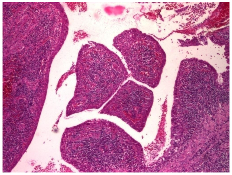
Background: This study presents a comprehensive analysis of cholesteatoma of the middle ear, focusing on its clinical presentation, diagnostic imaging, and treatment outcomes. Cholesteatomas are defined by the keratinized squamous epithelium within the middle ear, leading to significant bone erosion, often affecting the ossicular chain and surrounding structures. Methods: The study explores various mechanisms involved in cholesteatoma progression, including enzymatic lysis, inflammatory responses, and neurotrophic disturbances. The study conducted a retrospective clinical and statistical review of 580 patients over a 20-year period (2003-2023), highlighting the role of advanced imaging, including computed tomography (CT) and diffusion-weighted magnetic resonance imaging (DWI), in preoperative planning and postoperative follow-up. Results: Findings revealed that early detection and intervention are crucial in preventing severe complications such as intracranial infection and hearing loss. Surgical treatment primarily involved tympanoplasty and mastoidectomy, with a recurrence rate of 1.55% within two years. The study underscores the importance of integrating imaging advancements into clinical decision-making to enhance patient outcomes and suggests further investigation into molecular mechanisms underlying cholesteatoma progression and recurrence. Histopathological and microbiological analysis was performed to identify pathological patterns and microbial agents. Conclusions: The study highlights the importance of early diagnosis and intervention to prevent complications such as intracranial infections and permanent hearing loss, while also emphasizing the role of advanced imaging techniques in the management and long-term monitoring of cholesteatoma patients.

Keywords: CT; MRI; bone erosion; cholesteatoma; imaging; mastoidectomy; middle ear; radiography; tympanoplasty.


Conflict of interest statement

The authors declare no conflicts of interest.

Figures

Figure 1
Figure 1
A radiograph of the left petromastoid region in the temporo-tympanic (Schüller) incidence showed reduced transparency of most mastoid cells.
Figure 2
Figure 2
Reduced pneumatization of mastoid cells, with preservation of the bony septa between the cells.
Figure 3
Figure 3
Pneumatic cells with demineralized intercellular septa, with a faded, mottled, punctiform appearance, in places losing their individuality (accentuation of local vasomotor phenomena). There is no evidence of missing or defective bone structure.
Figure 4
Figure 4
Elements to reduce pneumatization and resorption of intercellular septa.
Figure 5
Figure 5
Small areas of osteolysis at the petrous portion, more evident anteriorly of the posterior margin of the temporal rock.
Figure 6
Figure 6
Small geode projected at the petrous region.
Figure 7
Figure 7
Postoperative control radiograph showing an area of osteolysis with clear, well-demarcated borders.
Figure 8
Figure 8
Osteocondensation of the petromastoid region with complete disappearance of pneumatization and evidence of the lateral venous sinus posterior to the posterior margin of the temporal rock.
Figure 9
Figure 9
Total loss of mastoid aeration accompanied by marked bone condensation changes (sclerosis).
Figure 10
Figure 10
Lack of mastoid air spaces in the pre- and retrosinusal cells, with visualization of the lateral venous sinus (band of increased transparency, 1–1.2 cm wide, with smooth borders, oblique from top to bottom, parallel to the posterior border of the rock).
Figure 11
Figure 11
Patient with diagnosis of right suppurative chronic polypous polypous otomastoiditis. CT examination in axial sections shows absence of mastoid cell pneumatization and presence of a tissue mass in the right external auditory canal.
Figure 12
Figure 12
Patient with diagnosis of chronic suppurative otomastoiditis, lateral venous sinus thrombosis. CT scan, axial section, bony window showing absence of pneumatization of left-sided mastoid cells, fluid retention in the area, and MR venous 2DTOF MRI venous 2DTOF sequence with absence of signal at the left-sided lateral venous sinus.
Figure 13
Figure 13
Patient with the diagnosis of chronic suppurative left exteriorized otomastoiditis. CT examination, in postcontrast CT axial sections, parenchymal window and bone window, with bone sequestration and fistulization in the subcutaneous soft parts.
Figure 14
Figure 14
Patient with diagnosis of chronic suppurative otomastoiditis, brain abscess. CT examination in axial sections, bone window, and parenchymal window, post-contrast, showed lack of pneumatization of left mastoid cells; in the brain substance adjacent to the posterior aspect of the left temporal rock, gas bubbles and diffuse and moderate contrast uptake.
Figure 15
Figure 15
Patient with the diagnosis of acute suppurative polypous exteriorized retroauricular acute polypoid otomastoiditis. CT examination in axial, bony window, and parenchymal sections revealed underdeveloped mastoid air spaces on the left side, tissue formation in the external auditory canal, and diffuse infiltration of the retroauricular soft tissues.
Figure 16
Figure 16
Patient with the diagnosis of right chronic suppurative polypous chronic polypous otomastoiditis, left chronic otomastoiditis, deviated nasal septum, and chronic hypertrophic rhinitis. CT examination, axial sections, bone, and parenchymal window—hypertrophy of bilateral middle nasal turbinates, accentuated on the left side; reduced pneumatization of mastoid cells on the left side; lack of air cell development in the mastoid cells; presence of effudion with increased densities in the right mastoid.
Figure 17
Figure 17
CT axial sections, bone, and parenchymal window showing right temporooccipital osteolysis and at the posterior aspect of the temporal rock on the right side, right temporooccipital epicranial collection and diffuse infiltration of the integument and retroauricular fat, right cerebellar subdural collection with subdural empyema appearance.
Figure 18
Figure 18
Left petromastoid region, Schüller’s incidence shows reduced left-sided mastoid pneumatization.
Figure 19
Figure 19
Axial CT sections, bone and parenchymal window—extensive area of osteolysis in the left temporal bone and external wall of the mastoid, absence of pneumatization of left-sided mastoid cells and reduced pneumatization of right-sided mastoid cells, epididymal collection, diffuse infiltrative appearance of the preauricular soft tissues.
Figure 20
Figure 20
Patient with the diagnosis of acute cholesteatomatousotomastoiditis, right temporal cerebritis, and neighboring meningeal reaction in MRI examination with axial and coronal sections native and postgadolinium, an area in frank hyperseminal at the right mastoid, with fluid appearance; postgadolinium, diffusely demarcated area, at the level of the brain parenchyma, right temporal lobe, and meningeal pathologic contrast uptake, right temporal.
Figure 21
Figure 21
Axial and coronal postgadolinium axial and coronal sections show right temporal cerebral abscesses and pathologic uptake of neighboring meningeal contrast.
Figure 22
Figure 22
Axial T2-weighted and T1-weighted MRI T2-weighted and T1-weighted MRI axial sections show hypersignal T2-weighted area, isosignal with the T1-weighted brain substance, located in the right middle ear; also, heterogeneous signal area is observed in the right mastoid, indicating the presence of superinfected fluid.
Figure 23
Figure 23
Patient with the diagnosis of chronic suppurative left retroauricular exteriorized chronic suppurative otomastoiditis, MRI examination with axial and coronal sections, with enlarged hyperseminal T2-weighted area in the middle ear and mastoid on the left side, hyposeminal T1-weighted.
Figure 24
Figure 24
Endoscopic examination of the right tympanic membrane with slight accentuation of the tympanic vascularization.
Figure 25
Figure 25
Cholesteatom, ob. ×10, col. HE.
Figure 26
Figure 26
Cholesteatom, ob. ×40, col. HE.
Figure 27
Figure 27
Polip, ob. ×4, col. HE.
Figure 28
Figure 28
Polip, ob. ×20, col. HE.
Figure 29
Figure 29
Tympanic membrane mucosa, epithelium with apocrine-like cells and chronic inflammatory infiltrate, ob. ×40, col. HE.
Figure 30
Figure 30
Tympanic membrane mucosa, epithelium with apocrine-like cells and chronic inflammatory infiltrate, ob. ×10, col. HE.
Figure 31
Figure 31
B lymphocytes in small amounts diffusely distributed, highlighted by IHC technique using CD20 atc, ob. ×100.
Figure 32
Figure 32
Much more numerous T lymphocytes arranged around blood vessels, ob. ×100.
Figure 33
Figure 33
T lymphocytes unevenly distributed, more abundant around blood vessels, granuloma-like appearance; T lymphocytes visualized by IHC technique using atc CD3, ob. ×100.
Figure 34
Figure 34
Much more numerous T lymphocytes arranged around blood vessels, ob. ×100.
Figure 35
Figure 35
T lymphocytes unevenly distributed, more abundant around blood vessels, granuloma-like appearance; T lymphocytes visualized by IHC technique using atc CD3, ob. ×100.

References

    1. Albera R., Canale A., Pimetto E., Lacilla M., Dagna F. Ossicular chain lesions in cholesteatoma. Acta Otorhinolaryngol. Ital. 2012;32:309–313. - PMC - PubMed
    1. Abramson M. Colagenolytic activity in middle ear cholesteatoma. Ann. Otol. 1969;78:112–124. - PubMed
    1. Brescia G., Frosolini A., Franz L., Daloiso A., Fantin F., Lovato A., Filippis C.d., Marioni G. Chronic Otitis Media in Patients with Chronic Rhinosinusitis: A Systematic Review. Medicina. 2023;59:123. doi: 10.3390/medicina59010123. - DOI - PMC - PubMed
    1. Sone M., Yoshida T., Naganawa S., Otake H., Kato K., Sano R., Teranishi M., Nakashima T. Comparison of computed tomography and magnetic resonance imaging for evaluation of cholesteatoma with labyrinthine fistulae. Laryngoscope. 2012;122:1121–1125. doi: 10.1002/lary.23204. - DOI - PubMed
    1. Tsuchida K., Takahashi M., Nakazawa T., Kurihara S., Yamamoto K., Yamamoto Y., Kojima H. Augmented Reality-Assisted Transcanal Endoscopic Ear Surgery for Middle Ear Cholesteatoma. J. Clin. Med. 2024;13:1780. doi: 10.3390/jcm13061780. - DOI - PMC - PubMed

LinkOut - more resources