. 2024 Nov 13;13(22):6826.
doi: 10.3390/jcm13226826.

MRI Spinal Cord Reconstruction Provides Insights into Mapping and Migration Following Percutaneous Epidural Stimulation Implantation in Spinal Cord Injury

Affiliations

MRI Spinal Cord Reconstruction Provides Insights into Mapping and Migration Following Percutaneous Epidural Stimulation Implantation in Spinal Cord Injury

Siddharth Venigalla et al. J Clin Med. .

Abstract

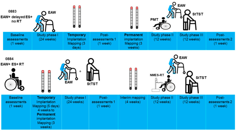
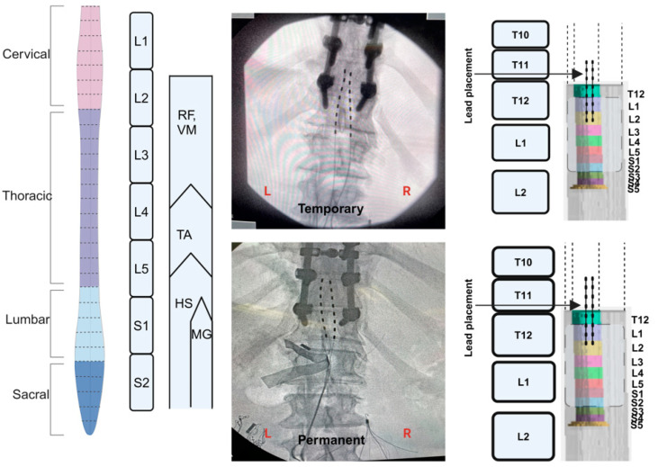
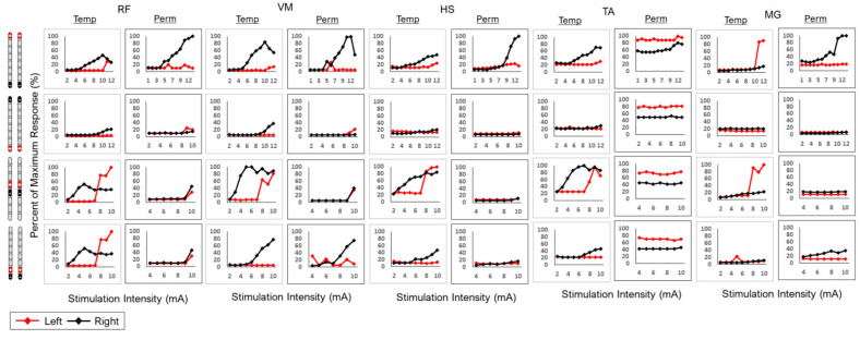
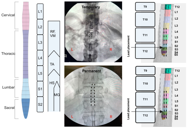
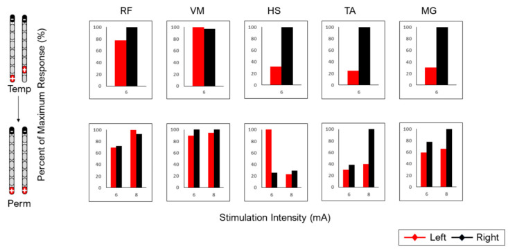
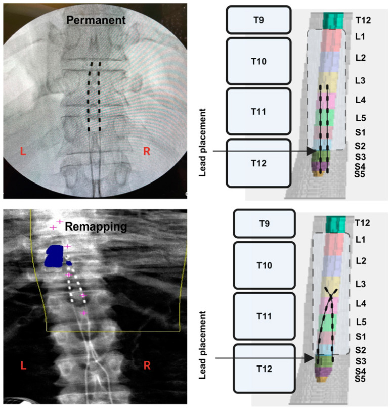
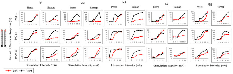
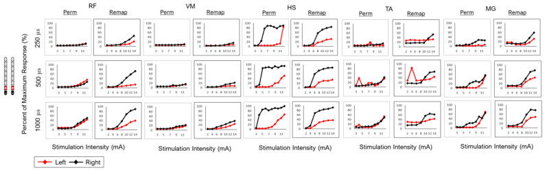
Background: Spinal cord epidural stimulation (SCES) has the potential to restore motor functions following spinal cord injury (SCI). Spinal cord mapping is a cornerstone step towards successfully configuring SCES to improve motor function, aiming to restore standing and stepping abilities in individuals with SCI. While some centers have advocated for the use of intraoperative mapping to anatomically target the spinal cord locomotor centers, this is a resource-intensive endeavor and may not be a feasible approach in all centers. Methods: Two participants underwent percutaneous SCES implantation as part of a clinical trial. Each participant underwent a temporary (1-week, two-lead) trial followed by a permanent, two-lead implantation. SCES configurations were matched between temporary and permanent mappings, and motor evoked potential in response to 2 Hz, for a duration of 250-1000 µs and with an amplitude of 1-14 mA, was measured using electromyography. T2 axial MRI images captured prior to implantation were used to retrospectively reconstruct the lumbosacral segments of the spinal cord. The effects of lead migration on mapping were further determined in one of the participants. Results: In both participants, there were recognized discrepancies in the recruitment curves of the motor evoked potentials across different muscle groups between temporary and permanent SCES mappings. These may be explained by retrospective MRI reconstruction of the spinal cord, which indicated that the percutaneous leads did not specifically target the entire L1-S2 segments in both participants. Minor lead migration appeared to have a minimal impact on spinal cord mapping outcomes in one of the participants but did dampen the motor activity of the hip and knee muscle groups. Conclusions: Temporary mapping coupled with MRI reconstruction has the potential to be considered as guidance for permanent implantation considering target activation of the spinal cord locomotor centers. Since lead migration may alter the synergistic coordination between different muscle groups and since lead migration of 1-2 contacts is expected and planned for in clinical practice, it can be better guided with proper spinal cord mapping and a diligent SCES lead trial beforehand.

Keywords: MRI; permanent mapping; spinal cord epidural stimulation (SCES); spinal cord injury; spinal cord reconstruction; temporary mapping.


Conflict of interest statement

The authors declare no conflict of interest.

Figures

Figure 1
Figure 1
Timeline of study phases for the two participants, 0883 and 0884. Both participants were randomized into either an exoskeletal-assisted walking (EAW) + 6 months delayed SCES + no resistance training (RT) group or EAW + SCES + RT. The timeline of the interventions was approximately 12 months separated by measurements at the 6-month period at baseline, post-assessment 1 and post-assessment 2 (1 week each; results are not reported). After baseline measurement, temporary implantation was performed, followed by 3–5 days of mapping. Following post-assessment 1, permanent implantation was performed and followed with mapping for another 3 weeks. The first 12 weeks following implantation included passive movement training (PMT; twice weekly) to balance the design with the other group (EAW + SCES + RT). For participant 0884, phase I of the study began with 6 months of training 3 days per week. After 24 weeks, one week of reassessing outcome measures (post-assessment 1) and four weeks of remapping were conducted to optimize standing and walking functions for the next phase of the study (interim mapping). Phase II of the study consisted of 12 weeks of the same training as in phase I, plus 2 days/week of neuromuscular electrical stimulation resistance training (NMES-RT) on alternate days to SCES + EAW and SCES + StTST. This was followed directly by phase III of the study, in which the participants continued the same training as in phase I but where NMES-RT was replaced by SCES + sit-to-stand practice with a walker for 12 weeks. In the final week, the outcome measures were reassessed (post-assessment 2).
Figure 2
Figure 2
Spinal cord model highlighting the lumbosacral innervation of the corresponding muscles. Fluoroscopy images following the conclusion of temporary and parament implantation in subject 0883. Retrospective 3D MRI reconstruction of the spinal cord and estimated placement of the percutaneous SCES leads during temporary and permanent mapping (subject 0883). The reconstructed spinal cord was based on axial MRI of the lumbosacral segments prior to implantation. The lower 3 contacts per lead covered the L1 and L2 segments. The measured distance from S5 to S2 was 1.5 cm. (Created in BioRender; Rehman, M. (2024); BioRender.com/d70p619 (accessed on 6 November 2024).).
Figure 3
Figure 3
Agreement between temporary and permanent mappings for different muscle groups (RF, VM, HS, TA and MG) in subject 0883. Four configurations were tested with two wide-field and two narrow-field configurations. All configurations were tested at 2 Hz with a pulse duration of 250 µs. Visual agreement was used to determine the similarity between the recruitment curves of the right-side temporary and right-side permanent or the left-side temporary and left-side permanent mappings.
Figure 4
Figure 4
Spinal cord model highlighting the lumbosacral innervation of the corresponding muscles. Fluoroscopy images following the conclusion of temporary and parament implantation in subject 0884. Retrospective 3D MRI reconstruction of the spinal cord and estimated placement of the percutaneous SCES leads during temporary and permanent mapping (subject 0884). The reconstructed spinal cord was based on axial MRI of the lumbosacral segments prior to implantation. The upper 6 contacts per lead covered the L3 and S2 segments. The measured distance from S5 to S2 was 1.5 cm. (Created in BioRender; Rehman, M. (2024); BioRender.com/w78b669 (accessed on 6 November 2024).).
Figure 5
Figure 5
Agreement between temporary and permanent mappings for different muscle groups (RF, VM, HS, TA and MG) in subject 0884. Only one amplitude (6 mA) was tested at a frequency of 2 Hz with a pulse duration of 500 µs during temporary implantation. Motor evoked responses were attenuated or remained silent at amplitudes of 1–4 mA following temporary implantation. Because of the retrospective nature of the work, amplitudes of 6 and 8 mA were chosen at a frequency of 2 Hz with a pulse duration of 500 µs during permanent mapping to determine the visual agreement compared to temporary mapping. The 8 mA amplitude was included because of the difference in anodal locations between temporary (+14) and permanent (+15) mappings of the chosen staggered configuration, which may have accounted for different motor evoked responses. The temporary and permanent motor evoked responses were similar for 4 muscle groups (RF, VM, TA and MG) and showed high visual agreement, except for HS. The HS muscle group showed a mirror response of the motor evoked potentials between the left and right sides when comparing permanent responses to temporary responses.
Figure 6
Figure 6
Lead migration in participant 0884. The tip of the left lead crossed over to the right lead at the L3 segment. Fluoroscopy images following the conclusion of permanent implantation in subject 0884. Lead migration was detected by dual-energy X-ray absorptiometry of the spine region. (Created in BioRender; Rehman, M. (2024); BioRender.com/t49c027 (accessed on 6 November 2024).).
Figure 7
Figure 7
Wide-field configuration with caudal cathode placement in subject 0884 following permanent implantation (perm mapping) and migration of the leads (remap: remapping) at different pulse durations (250, 500 and 1000 µs) in different muscle groups (RF, VM, HS, TA and MG). Visual agreement indicated attenuated responses in the left RF and HS following migration.
Figure 8
Figure 8
Caudal narrow-field configurations in subject 0884 following permanent implantation (perm mapping) and migration of the leads (remap: remapping) at different pulse durations (250, 500 and 1000 µs) in different muscle groups (RF, VM, HS, TA and MG). Visual agreement indicated attenuated responses in the left RF and HS following migration. The current had to be increased to 14 mA to achieve a similar response to what was achieved during permanent implantation. Results are listed in Table 2.

Similar articles

Cited by

References

    1. Anjum A., Yazid M.D., Fauzi Daud M., Idris J., Ng A.M.H., Selvi Naicker A., Ismail O.H.R., Athi Kumar R.K., Lokanathan Y. Spinal Cord Injury: Pathophysiology, Multimolecular Interactions, and Underlying Recovery Mechanisms. Int. J. Mol. Sci. 2020;21:7533. doi: 10.3390/ijms21207533. - DOI - PMC - PubMed
    1. Pang Q.M., Chen S.Y., Xu Q.J., Fu S.P., Yang Y.C., Zou W.H., Zhang M., Liu J., Wan W.H., Peng J.C., et al. Neuroinflammation and Scarring After Spinal Cord Injury: Therapeutic Roles of MSCs on Inflammation and Glial Scar. Front. Immunol. 2021;12:751021. doi: 10.3389/fimmu.2021.751021. - DOI - PMC - PubMed
    1. Guest J., Datta N., Jimsheleishvili G., Gater D.R., Jr. Pathophysiology, Classification and Comorbidities after Traumatic Spinal Cord Injury. J. Pers. Med. 2022;12:1126. doi: 10.3390/jpm12071126. - DOI - PMC - PubMed
    1. Diop M., Epstein D. A Systematic Review of the Impact of Spinal Cord Injury on Costs and Health-Related Quality of Life. PharmacoEconomics Open. 2024;8:793–808. doi: 10.1007/s41669-024-00517-3. - DOI - PMC - PubMed
    1. Turczyn P., Wojdasiewicz P., Poniatowski Ł.A., Purrahman D., Maślińska M., Żurek G., Romanowska-Próchnicka K., Żuk B., Kwiatkowska B., Piechowski-Jóźwiak B., et al. Omega-3 fatty acids in the treatment of spinal cord injury: Untapped potential for therapeutic intervention? Mol. Biol. Rep. 2022;49:10797–10809. doi: 10.1007/s11033-022-07762-x. - DOI - PubMed

LinkOut - more resources