

Evaluation of Dental Panoramic Radiographs by Artificial Intelligence Compared to Human Reference: A Diagnostic Accuracy Study
- PMID: 39598002
- PMCID: PMC11595016
- DOI: 10.3390/jcm13226859
Evaluation of Dental Panoramic Radiographs by Artificial Intelligence Compared to Human Reference: A Diagnostic Accuracy Study
Abstract
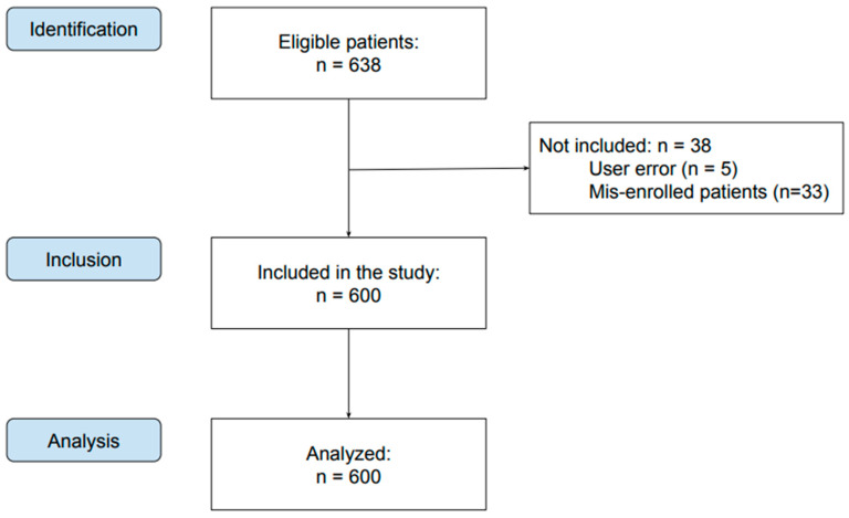
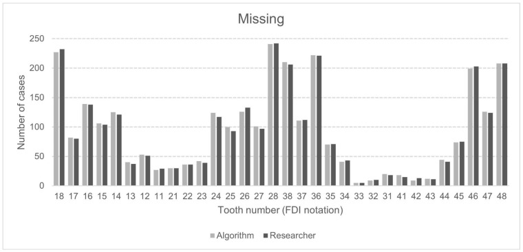
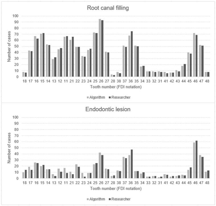
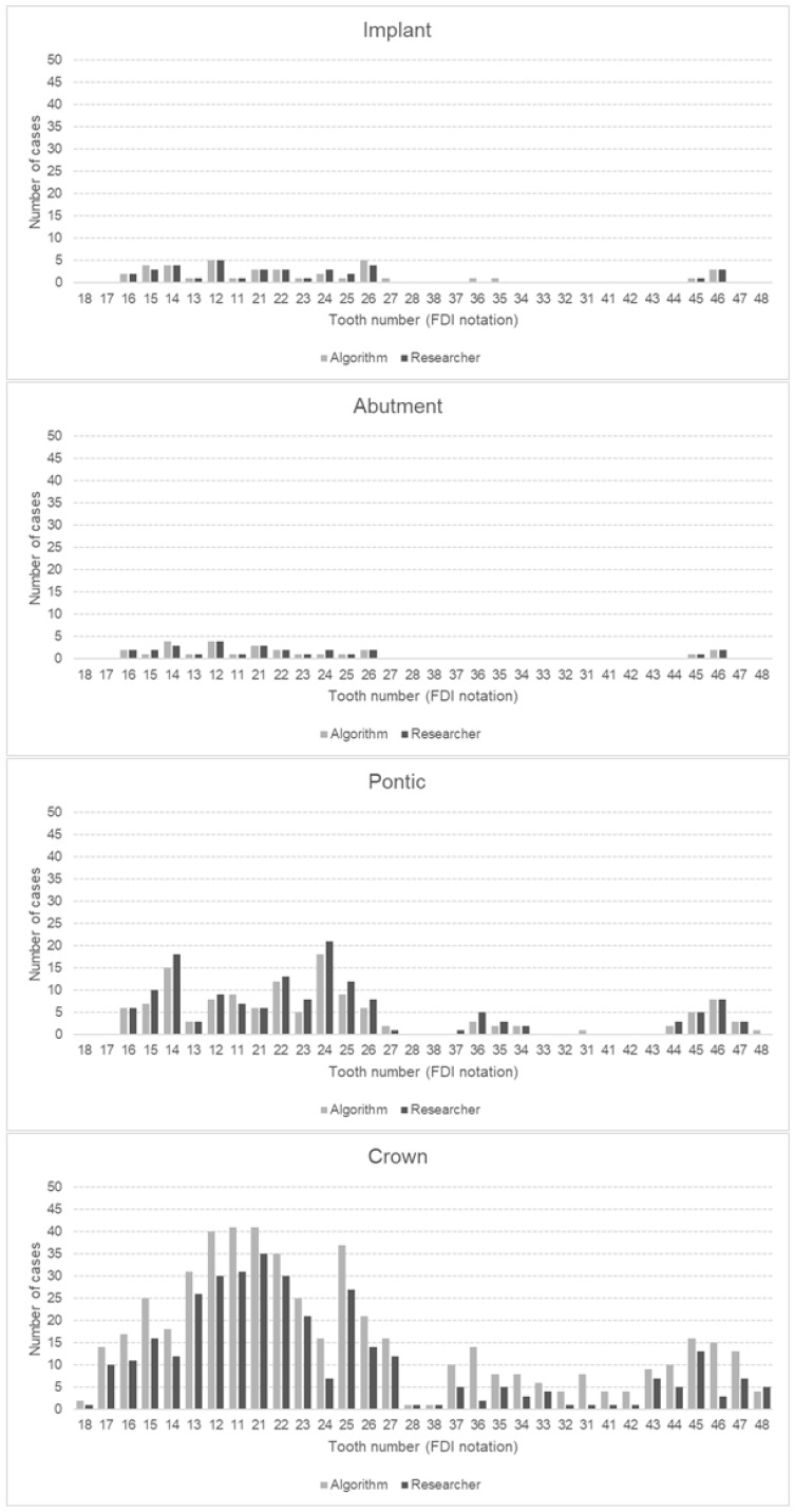
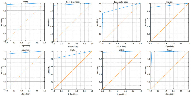
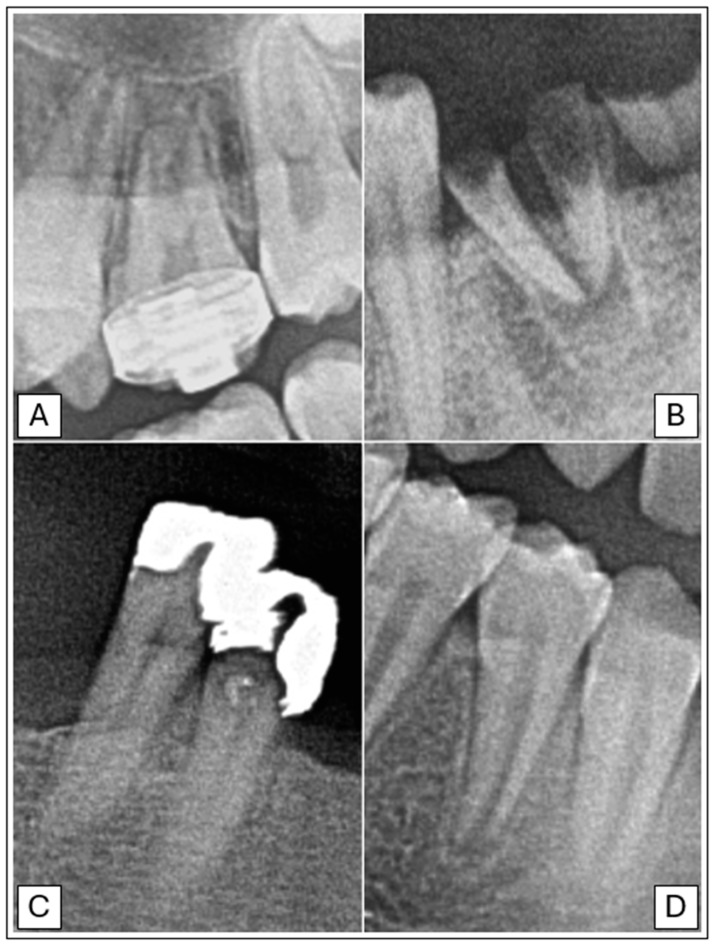
Background/Objectives: The role of artificial intelligence (AI) in dentistry is becoming increasingly significant, particularly in diagnosis and treatment planning. This study aimed to assess the sensitivity, specificity, accuracy, and precision of AI-driven software in analyzing dental panoramic radiographs (DPRs) in patients with permanent dentition. Methods: Out of 638 DPRs, 600 fulfilled the inclusion criteria. The radiographs were analyzed by AI software and two researchers. The following variables were assessed: (1) missing tooth, (2) root canal filling, (3) endodontic lesion, (4) implant, (5) abutment, (6) pontic, (7) crown, (8) and sound tooth. Results: The study revealed very high performance metrics for the AI algorithm in detecting missing teeth, root canal fillings, and implant abutment crowns, all greater than 90%. However, it demonstrated moderate sensitivity and precision in identifying endodontic lesions and the lowest precision (65.30%) in detecting crowns. Conclusions: AI software can be a valuable tool in clinical practice for diagnosis and treatment planning but may require additional verification by clinicians, especially for identifying endodontic lesions and crowns. Due to some limitations of the study, further research is recommended.
Keywords: artificial intelligence; automatic detection; diagnosis; panoramic radiograph.
Conflict of interest statement
The authors declare no conflicts of interest.
Figures
Similar articles
-
Artificial Intelligence (AI) Assessment of Pediatric Dental Panoramic Radiographs (DPRs): A Clinical Study.Pediatr Rep. 2024 Sep 11;16(3):794-805. doi: 10.3390/pediatric16030067. Pediatr Rep. 2024. PMID: 39311330 Free PMC article.
-
Oral Health Status and Treatment Needs Based on Artificial Intelligence (AI) Dental Panoramic Radiograph (DPR) Analysis: A Cross-Sectional Study.J Clin Med. 2024 Jun 25;13(13):3686. doi: 10.3390/jcm13133686. J Clin Med. 2024. PMID: 38999252 Free PMC article.
-
Assessment of the Diagnostic Accuracy of Artificial Intelligence Software in Identifying Common Periodontal and Restorative Dental Conditions (Marginal Bone Loss, Periapical Lesion, Crown, Restoration, Dental Caries) in Intraoral Periapical Radiographs.Diagnostics (Basel). 2025 Jun 4;15(11):1432. doi: 10.3390/diagnostics15111432. Diagnostics (Basel). 2025. PMID: 40507004 Free PMC article.
-
Applications of artificial intelligence in the analysis of dental panoramic radiographs: an overview of systematic reviews.Dentomaxillofac Radiol. 2023 Oct;52(7):20230284. doi: 10.1259/dmfr.20230284. Epub 2023 Sep 4. Dentomaxillofac Radiol. 2023. PMID: 37665008 Free PMC article. Review.
-
Artificial intelligence in dentistry and dental biomaterials.Front Dent Med. 2024 Dec 23;5:1525505. doi: 10.3389/fdmed.2024.1525505. eCollection 2024. Front Dent Med. 2024. PMID: 39917699 Free PMC article. Review.
Cited by
-
Artificial Intelligence and Dentomaxillofacial Radiology Education: Innovations and Perspectives.Dent J (Basel). 2025 May 29;13(6):245. doi: 10.3390/dj13060245. Dent J (Basel). 2025. PMID: 40559148 Free PMC article. Review.
-
The Validation of an Artificial Intelligence-Based Software for the Detection and Numbering of Primary Teeth on Panoramic Radiographs.Diagnostics (Basel). 2025 Jun 11;15(12):1489. doi: 10.3390/diagnostics15121489. Diagnostics (Basel). 2025. PMID: 40564810 Free PMC article.
-
AI Efficiency in Dentistry: Comparing Artificial Intelligence Systems with Human Practitioners in Assessing Several Periodontal Parameters.Medicina (Kaunas). 2025 Mar 23;61(4):572. doi: 10.3390/medicina61040572. Medicina (Kaunas). 2025. PMID: 40282863 Free PMC article.
-
Dental practitioners versus artificial intelligence software in assessing alveolar bone loss using intraoral radiographs.J Taibah Univ Med Sci. 2025 May 9;20(3):272-279. doi: 10.1016/j.jtumed.2025.04.001. eCollection 2025 Jun. J Taibah Univ Med Sci. 2025. PMID: 40476084 Free PMC article.
References
-
- Różyło-Kalinowska I. Panoramic radiography in dentistry. Clin. Dent. Rev. 2021;5:26. doi: 10.1007/s41894-021-00111-4. - DOI
-
- Farman A.G. Panoramic Radiology. Seminars on Maxillofacial Imaging and Interpretation. Springer; Berlin/Heidelberg, Germany: New York, NY, USA: 2007.
LinkOut - more resources
Full Text Sources
