. 2024 Oct 28;17(11):1443.
doi: 10.3390/ph17111443.

Effect of Hypoxia on Siglec-7 and Siglec-9 Receptors and Sialoglycan Ligands and Impact of Their Targeting on NK Cell Cytotoxicity

Affiliations

Effect of Hypoxia on Siglec-7 and Siglec-9 Receptors and Sialoglycan Ligands and Impact of Their Targeting on NK Cell Cytotoxicity

Husam Nawafleh et al. Pharmaceuticals (Basel). .

Abstract

Background/objectives: Tumor microenvironmental hypoxia is an established hallmark of solid tumors. It significantly contributes to tumor aggressiveness and therapy resistance and has been reported to affect the balance of activating/inhibitory surface receptors' expression and activity on NK cells. In the current study, we investigated the impact of hypoxia on the surface expression of Siglec-7 and Siglec-9 (Sig-7/9) and their ligands in NK cells and tumor target cells. The functional consequence of Siglec blockage using nanoparticles specifically designed to target and block Sig-7/9 receptors on NK cell cytotoxicity was elucidated.

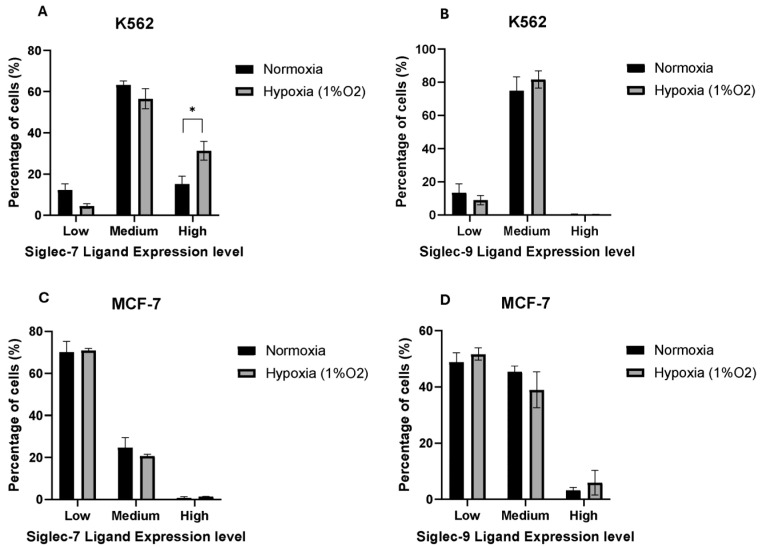
Methods: CD56⁺ CD3- NK cells were isolated from PBMCs along with an NK-92 clone and used as effector cells, while MCF-7 and K562 served as target cells. All cells were incubated under normoxic or hypoxic conditions for 24 h. To assess Siglec-7 and Siglec-9 receptor expression, U937, NK-92, and primary NK cells were stained with PE-labeled antibodies against CD328 Siglec-7/9. Interactions between Siglec-7/9 and their sialylated ligands, along with their functional impact on NK cell activity, were evaluated using polymeric nanoparticles coated with a sialic acid mimetic. Immunological synapse formation and live-cell imaging were performed with a ZEISS LSM 800 with Airyscan at 10× magnification for 24 h.

Results: Our data indicate that hypoxia had no effect on the expression of Siglec-7/9 receptors by NK cells. In contrast, hypoxic stress resulted in an increase in Siglec-7 sialoglycan ligand expression by a sub-population of NK target cells. Using polymeric nanoparticles coated with a sialic acid mimetic that binds both Siglec-7 and -9 (Sig-7/9 NP), we demonstrated that incubation of these nanoparticles with NK cells resulted in increased immunological synapse formation, granzyme B accumulation, and killing of NK target cells. These studies indicate that hypoxic stress may have an impact on NK cell-based therapies and highlight the need to consider the hypoxic microenvironment for tumor-specific glycosylation.

Conclusions: Our findings point to the role of Siglec-sialylated glycan interactions in hypoxic stress-induced NK cell dysfunction and recommend the potential integration of the manipulation of this axis through the targeting of Siglecs in future cancer immunotherapy strategies.

Keywords: NK cells; hypoxia; immune synapses; nanoparticles; sialoglycan ligands; siglec receptors.


Conflict of interest statement

The authors (A.K., L.H., M.G., D.K. and M.T.) are full-time employees of Aviceda Therapeutics. The remaining authors (H.N., N.Z., M.K.G., C.J.S. and S.C.) declare that the research was conducted in the absence of any commercial or financial relationships that could be construed as a potential conflict of interest.

Figures

Figure 1
Figure 1
The effect of hypoxia on the surface expression of Siglec-7 and Siglec-9 receptors on immune cells. Histograms of one representative experiment show the effect of hypoxia (1% oxygen) for 24 h in comparison with normoxia (21% oxygen) on the surface expression of Siglec-7 and Siglec-9. (A,B) NK-92; (C,D) primary NKs; (E,F) U937 monocytes cell line. The bar charts represent the mean of the Median Fluorescent Intensity (MFI) of Siglec-7 Receptor (G) and Siglec-9 Receptor (H) on the surface of the indicated effector cells. U937 monocytes were used as a positive control while unstained samples for each experiment were used as negative controls. Error bars represent SEM of three independent experiments.
Figure 2
Figure 2
The effect of hypoxia on the surface expression of Siglec-7 and Siglec-9 ligands on tumor cell lines. The bar charts represent the Median Fluorescent Intensity (MFI) of (A) Siglec-7 ligand and (B) Siglec-9 ligand on the surface of K562 and MCF-7 cell lines in hypoxia and normoxia. Error bars represent SEM of three independent experiments. (**) p value ≤ 0.01 based on unpaired t-test.
Figure 3
Figure 3
The effect of hypoxia on the regulation of Siglec-7 and Siglec-9 ligands on the surface of the K562 and MCF-7 cell lines. The bar charts show the percentages of K562 cells that express Siglec-7 ligand (A) and Siglec-9 ligand (B), as well as MCF-7 cells that express Siglec-7 ligand (C) and Siglec-9 ligand (D) at different levels: low, medium, and high expression levels, as assessed by flow cytometry. Error bars represent SEM of three independent experiments. (*) p value ≤ 0.05 based on unpaired t-test.
Figure 4
Figure 4
Immunological synapse formation. (A) Inhibition of Siglec-7/Fc chimera and Siglec-9/Fc chimera binding to K562 cells by Sig-7/9 NPs. Representative histogram shown from three independent experiments. (B) Confocal imaging of the accumulation of granzyme B, actin, and phospho-tyrosine between Sig-7/9 NPs and Control NPs treated and non-treated peripheral NK and MCF-7 cells representing immunological synapse formation and/or recognition. Scale bar = 10 µm. (C) Quantification of the treated and non-treated peripheral NK cells recognizing and/or forming an immunological synapse with MCF-7. Error bars represent SEM of three independent experiments. Statistical significance was determined using two-way ANOVA. (***) p value ≤ 0.001; (****) p ≤ 0.0001.
Figure 5
Figure 5
The effect of nanoparticles on primary NK-mediated cytotoxicity. Target tumor cell lines (A) K562 and (B) MCF-7 were co-cultured with treated primary NK cells and assessed using LDH cytotoxicity assay. Error bars represent SEM of three independent experiments. Statistical significance was determined using two-way ANOVA. (*) p value ≤ 0.05; (**) p value ≤ 0.01.
Figure 6
Figure 6
Live-cell imaging of peripheral NK cytotoxicity treated with Sig-7/9 NPs, Control NPs and non-treated peripheral NK cytotoxicity on MCF-7 cells. MCF-7 cells labeled with CMFDA green co-cultured with primary NK labeled with CMTMR orange. Dead cells shown in red. (A) Representative confocal images taken at different time points (0, 4 and 8 h). Scale bar = 100 µm. Orange arrows are pointing at the pNK cells forming immunological synapses with the target cells. (B) The percentages of killed MCF-7 cells by the treated and non-treated peripheral NK. Error bars represent SEM of three independent experiments. Statistical significance was determined using two-way ANOVA. (*) p value ≤ 0.05.

References

    1. Cha J.-H., Chan L.-C., Song M.S., Hung M.-C. New Approaches on Cancer Immunotherapy. Cold Spring Harb. Perspect. Med. 2019;10:a036863. doi: 10.1101/cshperspect.a036863. - DOI - PMC - PubMed
    1. Gajewski T.F., Schreiber H., Fu Y.-X. Innate and adaptive immune cells in the tumor microenvironment. Nat. Immunol. 2013;14:1014–1022. doi: 10.1038/ni.2703. - DOI - PMC - PubMed
    1. Horn L.A., Fousek K., Palena C. Tumor Plasticity and Resistance to Immunotherapy. Trends Cancer. 2020;6:432–441. doi: 10.1016/j.trecan.2020.02.001. - DOI - PMC - PubMed
    1. Hasmim M., Messai Y., Ziani L., Thiery J., Bouhris J.-H., Noman M.Z., Chouaib S. Critical Role of Tumor Microenvironment in Shaping NK Cell Functions: Implication of Hypoxic Stress. Front. Immunol. 2015;6:482. doi: 10.3389/fimmu.2015.00482. - DOI - PMC - PubMed
    1. Li Y., Zhao L., Li X.-F. Hypoxia and the Tumor Microenvironment. Technol. Cancer Res. Treat. 2021;20:15330338211036304. doi: 10.1177/15330338211036304. - DOI - PMC - PubMed

LinkOut - more resources