. 2024 Nov 20;29(22):5476.
doi: 10.3390/molecules29225476.

Mutational Analysis of Substrate Recognition in Trypsin-like Protease Cocoonase: Protein Memory Induced by Alterations in Substrate-Binding Site

Affiliations

Mutational Analysis of Substrate Recognition in Trypsin-like Protease Cocoonase: Protein Memory Induced by Alterations in Substrate-Binding Site

Nana Sakata et al. Molecules. .

Abstract

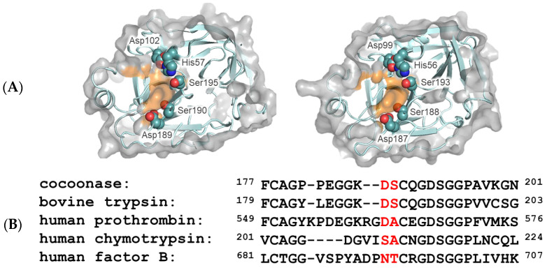
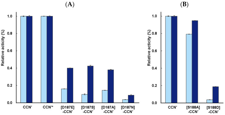
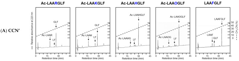
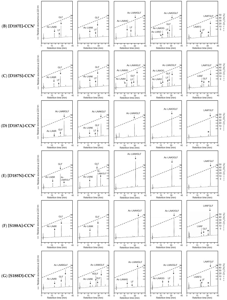
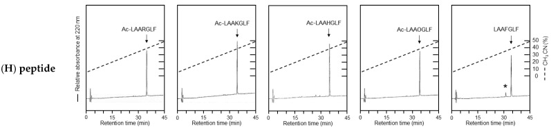
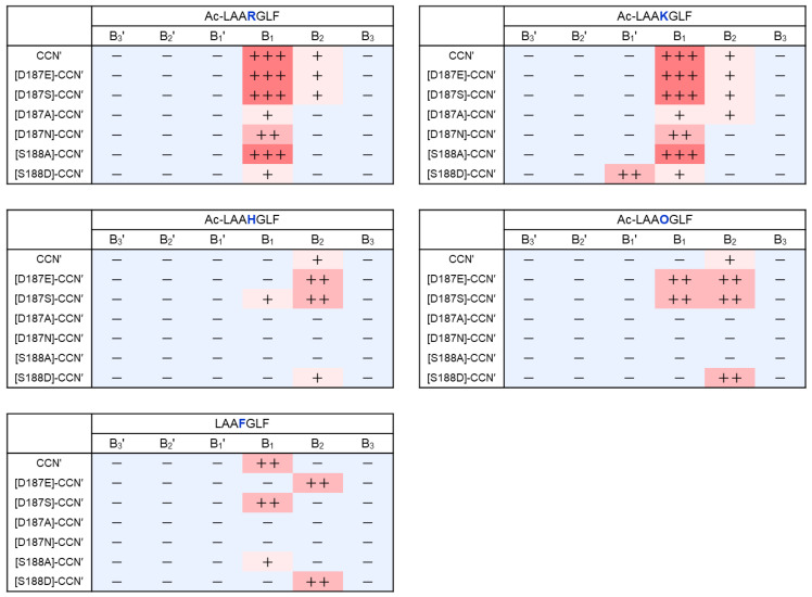
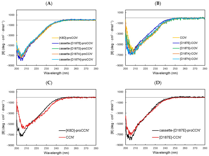
To investigate the substrate recognition mechanism of trypsin-like protease cocoonase (CCN), mutational analyses were conducted at key substrate recognition sites, Asp187 and Ser188, and their effects on substrate specificity and enzymatic activity were evaluated. Mutants with the Asp187 substitution exhibited a significant reduction in catalytic activity compared with the wild-type enzyme, whereas the Ser188 mutants displayed a comparatively minor effect on activity. This indicates that Asp187 plays a crucial role in catalytic function, whereas Ser188 serves a complementary role in substrate recognition. Interestingly, the substitution of the Asp187 to Glu or Ser caused novel substrate specificities, resulting in the recognition of Orn and His residues. In addition, when Asp187 and Ser188 were substituted with acidic residues (Glu or Asp), both the precursor proCCN and mature CCN proteins retained highly similar secondary and tertiary structures. This reveals that the structural characteristics of precursor proteins are maintained in the mature proteins, potentially influencing substrate recognition and catalytic function. These findings suggest that the pro-regions of these mutants interact much more tightly with the mature enzyme than in the wild-type CCN. These results provide fruitful insights into the structural determinants governing substrate recognition in enzyme variants.

Keywords: chaperone; chymotrypsin; cocoonase; folding; substrate binding.


Conflict of interest statement

The authors declare no conflicts of interest.

Figures

Figure 1
Figure 1
Molecular modeling of trypsin and cocoonase and their substrate-binding regions. (A) The molecular structures of the substrate-binding regions of trypsin (PDB: 5T3H, left) and Bombyx mori cocoonase (calculated using AlphaFold2, right). Surfaces of the amino acid residues at the S1 site are highlighted in orange. The catalytic triad and substrate-binding residues are indicated. (B) Alignment of the amino acid sequences of the substrate-binding regions of trypsin-like proteases. The predominant substrate-binding residues are indicated by red letters.
Figure 2
Figure 2
Casein zymography of the cocoonase (CCN) proteins mutated at substrate-binding sites, Asp187 (A) and Ser188 (B). (A) Lanes 1–5 represent the CCN′ and [D187E]-, [D187A]-, [D187N]-, and [D187S]-CCN′ proteins, respectively. (B) Lanes 1–5 represent the [S188A]-, [S188D]-, [S188E]-, [D187E,S188D]-, and [D187E,S188E]-CCN′ proteins, respectively. The CCN′ protein at lane 1 was derived from the [K8D]-proCCN′ protein. “M” represents marker proteins.
Figure 3
Figure 3
Casein assay of the CCN′ proteins mutated at putative substrate-binding sites, (A) Asp187 and (B) Ser188. The reaction times were 3 h (light blue) and 16 h (blue). Enzyme assays were conducted in triplicate (n = 3). The enzymatic activity of the CCN′ protein was normalized to the proteolytic activity of 1.0. * This CCN′ protein was derived from cassette-proCCN′.
Figure 4
Figure 4
HPLC profiles of the peptide substrates treated with the CCN′ mutant proteins. The peptide fragments were analyzed by amino acid analysis. Their identified sequences are indicated in the profiles. (A) The CCN′ protein was derived from [K8D]-proCCN′. (BG) represent [D187E]-, [D187S]-, [D187A]-, [D187N]-, [S188A]-, and [S188D]-CCN′. (H) The peptide substrates were allowed to stand for 3 days at 37 °C without the mutant proteins. There were no significant digests in the absence of the mutant enzymes. Asterisks (*) indicate impurities derived from the LAAFGLF peptide.
Figure 4
Figure 4
HPLC profiles of the peptide substrates treated with the CCN′ mutant proteins. The peptide fragments were analyzed by amino acid analysis. Their identified sequences are indicated in the profiles. (A) The CCN′ protein was derived from [K8D]-proCCN′. (BG) represent [D187E]-, [D187S]-, [D187A]-, [D187N]-, [S188A]-, and [S188D]-CCN′. (H) The peptide substrates were allowed to stand for 3 days at 37 °C without the mutant proteins. There were no significant digests in the absence of the mutant enzymes. Asterisks (*) indicate impurities derived from the LAAFGLF peptide.
Figure 5
Figure 5
Heat map of substrate specificity of the CCN′ mutant proteins. The cleaved sites identified by the mutant proteins are summarized, with the relative strength of cleavage represented using the number of “+” symbols. +++: strong, ++: moderate, +: weak, −: not detected.
Figure 6
Figure 6
CD spectra of a series of the proCCN′ (A) and CCN′ (B) mutant proteins. (A) Precursor proteins, (B) mature proteins, (C) [K8D]-proCCN′ and CCN′, and (D) cassette-[D187E]-proCCN′ and [D187E]-CCN′.

References

    1. Hedstrom L. Serine protease mechanism and specificity. Chem. Rev. 2002;102:4501–4524. doi: 10.1021/cr000033x. - DOI - PubMed
    1. Huber R., Bode W. Structural basis of the activation and action of trypsin. Acc. Chem. Res. 1978;11:114–122. doi: 10.1021/ar50123a006. - DOI
    1. Evnin L.B., Vasquez J.R., Craik C.S. Substrate specificity of trypsin investigated by using a genetic selection. Proc. Natl. Acad. Sci. USA. 1990;87:6659–6663. doi: 10.1073/pnas.87.17.6659. - DOI - PMC - PubMed
    1. Briand L., Chobert J.M., Gantier R., Declerck N., Tran V., Leonil J., Molle D., Haertle T. Impact of the lysine-188 and aspartic acid-189 inversion on activity of trypsin. FEBS Lett. 1999;442:43–47. doi: 10.1016/S0014-5793(98)01611-1. - DOI - PubMed
    1. Perona J.J., Craik C.S. Structural basis of substrate specificity in the serine proteases. Protein Sci. 1995;4:337–360. doi: 10.1002/pro.5560040301. - DOI - PMC - PubMed

Substances

LinkOut - more resources